







Executive Overview
For decades, evolutionary biologists have mapped the trajectory of vertebrate development with a sense of linear confidence. From swimming precursors to terrestrial pioneers, the general consensus held that the sophisticated optical systems of modern humans, birds, reptiles, and fish evolved through the steady refinement of an original, dual-eyed blueprint.
Groundbreaking research emerging from Lund University and the University of Sussex shatters this long-held paradigm. According to a comprehensive comparative analysis of light-detecting cells, our earliest vertebrate ancestors passed through an astonishing "cyclops-like" phase nearly 600 million years ago. During this period, these tiny, worm-like marine organisms relied on a single, centrally located median eye positioned on top of the head.
This revelation does more than merely add a bizarre footnote to the tree of life; it fundamentally rewrites the evolutionary history of the eye and the brain. The study indicates that the vertebrate visual system—including the high-definition paired eyes we use to navigate the world today—is the product of an unexpected evolutionary detour. Even more remarkably, the remnants of this ancient median eye do not belong exclusively to the fossil record. They persist inside the modern human brain as the pineal gland, the enigmatic endocrine organ responsible for orchestrating our sleep-wake cycles and circadian rhythms.
By bridging paleontology, developmental biology, and comparative anatomy, this research solves one of the most persistent anomalies in evolutionary science: why vertebrate eyes are structurally inverted compared to the camera-like eyes of invertebrates such as squid and insects. As the scientific community digests these findings, our understanding of human anatomy is forced to look inward—revealing that every blink, dream, and circadian shift is tied to the legacy of a prehistoric, one-eyed ocean dweller.
Detailed Chronology: From the Ediacaran Seas to the Modern Brain
To comprehend how modern humanity inherited its visual architecture from a prehistoric cyclops, evolutionary biologists must retrace a timeline spanning six hundred million years. This journey begins in the shallow, primordial seas of the Ediacaran period, winds through a radical lifestyle shift, and culminates in the complex neuroanatomy of the modern vertebrate skull.
Phase 1: The Dual-Eyed Dawn and the Stationary Retreat
Long before backbones, jaws, or skulls emerged, the common ancestor of all vertebrates was a soft-bodied, worm-like creature drifting or burrowing in ancient marine environments. Early in this lineage’s history, the organism possessed paired light-sensitive regions or rudimentary eyes. In the animal kingdom, paired eyes are an evolutionary default for mobile organisms; they provide essential stereoscopic input, allowing free-swimming creatures to judge distance, calculate trajectories, and detect approaching predators from multiple angles.
However, evolutionary pressures are rarely static. As the lineage adapted to a remarkably sedentary lifestyle—anchoring itself to the ocean floor and filtering passing seawater for plankton—the adaptive value of paired vision plummeted. For an organism that no longer hunted, chased, or actively navigated dynamic landscapes, maintaining complex, laterally positioned eyes was an unnecessary metabolic drain.
Over countless generations, natural selection favored the reduction and eventual loss of these paired visual structures. Yet, total blindness was not an option in a sunlit ocean where vertical orientation and day-night cycles dictated survival. While the lateral eyes faded into evolutionary obscurity, a cluster of light-sensitive cells situated precisely in the middle of the creature’s head remained.
Phase 2: The Reign of the Median Cyclops
With its lateral eyes gone, the organism entered its "cyclopean" epoch. A single, primitive median eye dominated the top of its head.
Biomechanical and ecological reconstructions suggest this central eye was incapable of forming crisp, high-resolution images. Instead, it served as a vital environmental compass. By detecting the direction of down-welling sunlight, the median eye allowed the creature to distinguish day from night and determine upward from downward—a crucial capability for filtering plankton and avoiding surface hazards.
Crucially, this median eye was structurally integrated with the organism’s developing neural tissues. It established baseline biological rhythms tied to solar cycles, embedding light-response mechanisms deep within the proto-brain.
Phase 3: The Great Active Re-emergence
Millions of years passed. The ecological landscape shifted, and descendants of these stationary filter-feeders abandoned their sedentary existence, returning to an active, swimming lifestyle.
This return to mobility instantly resurrected the evolutionary demand for advanced vision. To hunt, evade predators, and navigate complex marine terrains, the organism once again needed directional, image-forming eyes. Rather than reinventing vision from scratch or re-evolving the lost lateral structures, evolution took an unprecedented shortcut.
The organism repurposed portions of the ancient median eye. Through radical developmental shifts, regionalized cells from this central visual structure expanded, migrated, and adapted, eventually giving rise to a brand-new pair of image-forming eyes. This unusual recycling process explains a foundational divergence in the animal kingdom: why vertebrate retinas develop outward from the brain tissue itself, while the eyes of invertebrates like insects and mollusks originate from the surface ectoderm (skin) on the sides of the head.
Supporting Context & Metrics: Unraveling the Vertebrate Anomaly
To validate a hypothesis this disruptive, the research teams at Lund University and the University of Sussex had to assemble a massive evidentiary framework. For generations, comparative anatomists have puzzled over the structural "imperfection" of the vertebrate retina.
The Retinal Inversion Paradox
In cephalopods (such as octopuses and squid) and arthropods (such as insects), the retina is constructed logically. Light-gathering photoreceptor cells point directly toward the light source, and nerve fibers route neatly behind them, exiting the eye without obstructing incoming photons.
Vertebrates, by contrast, possess an "inverted" retina. The light-sensitive cells are buried at the very back of the tissue layer. Incoming light must pass through a dense meshwork of blood vessels and transparent nerve fibers before it finally strikes the photoreceptors. For years, creationists and evolutionary critics alike pointed to this structural quirk as an inefficient design flaw.
The new research resolves this paradox completely. The vertebrate retina’s inverted architecture is not a flaw; it is the indelible fingerprint of its origin. Because vertebrate eyes evolved from brain tissue (specifically, outgrowths of the central median eye structure) rather than folding inward from the surface skin, the neural wiring and blood supply were inherently positioned in front of the photoreceptors.
Decoding Neural Circuits
Furthermore, this discovery sheds light on the origins of the complex neural circuits embedded within the retina. Before visual signals ever reach the cerebral cortex, the vertebrate retina performs sophisticated computations—analyzing edge contrast, motion vectors, and luminance gradients.
By tracing these pathways back to the median eye of a 600-million-year-old worm-like ancestor, scientists now understand that retinal computation is an ancient inheritance. The foundational neural networks designed to process light signals in a single, primitive median eye were duplicated and expanded when the paired eyes formed, laying the neurological groundwork for the hyper-complex visual cortex found in mammals today.
Official Statements: Rethinking Sensory Evolution
The implications of this study have sent shockwaves through the fields of evolutionary biology and neuroanatomy. Dan-E Nilsson, professor emeritus in sensory biology at Lund University and a leading voice in the research, emphasizes the profound shift in perspective forced by these findings.
"The results are a surprise. They turn our understanding of the evolution of the eye and the brain upside down," notes Professor Nilsson.
The realization that vertebrate optical systems trace their ancestry through a single, central bottleneck challenges textbook assumptions about how complex organs develop. Rather than a steady accumulation of progressive improvements, vertebrate vision emerged from a sequence of loss, retention, and radical repurposing.
Addressing the structural uniqueness of human and animal sight, Nilsson highlights how the new model finally solves a decades-old comparative biology debate:
"Now we finally understand why the eyes of vertebrates differ so radically from the eyes of all other animal groups, such as insects and squid. The film of our eyes—the retina—developed from the brain, whereas the eyes of insects and squid originate in the skin on the sides of the head."
Perhaps most poetically, Nilsson points to the persistent ghost of our prehistoric ancestor resting quietly within our skulls:
"For the first time, we now also understand the origin of the neural circuits that analyze the image in our retina… It’s mind-boggling that our pineal gland’s ability to regulate our sleep according to light stems from the cyclopean median eye of a distant ancestor 600 million years ago."
Future Outlook: The Pineal Gland and Modern Medicine
As the academic paper gains traction across international research institutions, the ripple effects extend far beyond evolutionary theory into neurobiology, chronobiology, and clinical medicine.
The Pineal Gland as a Living Relic
The identification of the pineal gland as the direct evolutionary descendant of the median cyclopean eye opens new avenues for understanding circadian biology. The pineal gland—often historically dubbed the "third eye" by philosophers like René Descartes—is indeed a third eye in the most literal, evolutionary sense possible.
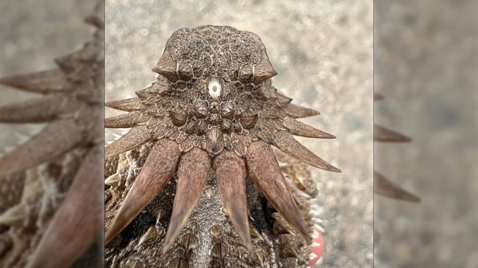
While human pineal glands do not form images, they retain core biochemical machinery dedicated to light responsiveness. In mammals, light information captured by the paired modern eyes travels along the retinohypothalamic tract to the suprachiasmatic nucleus, which in turn commands the pineal gland to suppress or release melatonin. In many lower vertebrates (such as certain lizards, fish, and amphibians), the pineal organ remains directly photosensitive, lying just beneath a thin patch of skull and skin.
Clinical Implications for Circadian Disorders
Understanding the deep evolutionary roots of the pineal gland provides fresh context for modern sleep medicine. In an era defined by artificial blue light, screen time, and chronic circadian disruption, humans frequently experience friction between their ancient neurobiology and modern lifestyles.
The pineal gland’s vulnerability to modern lighting environments is a direct consequence of its ancient mandate: to synchronize internal physiology with planetary light-dark cycles. By mapping the precise genetic and developmental pathways through which the median eye transformed into an endocrine regulator, researchers hope to design targeted therapies for circadian rhythm disorders, seasonal affective disorder (SAD), and age-related sleep fragmentation.
The Broader Scientific Horizon
Moving forward, evolutionary developmental biologists (evo-devo) plan to sequence and compare the regulatory genes controlling light-sensitive cell differentiation across a wider array of marine invertebrates. By isolating the specific genetic switches that allowed brain tissue to transform into optical structures, scientists may soon unlock regenerative protocols for human retinal diseases. If evolution could successfully repurpose a primitive median eye into the sophisticated visual systems of modern vertebrates, unlocking those same molecular pathways could eventually allow modern medicine to repair damaged retinas and restore sight to the blind.
Ultimately, this study serves as a humbling reminder of biological history. When humanity looks up at the stars or gazes into a mirror, we are peering through lenses forged by an ancient, single-eyed creature drifting in the quiet, sun-dappled oceans of a prehistoric world.