









Executive Overview
In what biomedical engineers and neuroscientists are heralding as a monumental leap forward for bioelectronic medicine, researchers at Northwell Health’s Feinstein Institutes for Medical Research have completed the world’s first comprehensive, high-resolution anatomical map of the human vagus nerve.
Spanning the brainstem to the abdomen, the vagus nerve serves as the body’s primary information superhighway, regulating involuntary processes as diverse as heart rate, respiration, digestion, and systemic immune responses. Despite its critical role in human health and its long-established status as a target for neuromodulation therapies, the internal architecture of the nerve—specifically, how hundreds of thousands of microscopic nerve fibers are spatially organized within its bundles—has remained largely an anatomical enigma.
Driven by a $6.7 million grant from the National Institutes of Health (NIH), a multidisciplinary team analyzed 60 vagus nerves from 30 human donors over a rigorous three-year period. By deploying cutting-edge micro-computed tomography (microCT), high-resolution ultrasound, fluorescent protein markers, and advanced immunohistochemistry, the research team successfully rendered an unprecedented three-dimensional reconstruction of the nerve’s complex internal wiring.
This newly released open-source dataset, now publicly accessible via the Stimulating Peripheral Activity to Relieve Conditions (SPARC) portal, promises to fundamentally alter how medical device engineers design next-generation neurostimulation therapies. By offering an unprecedented look at how distinct fiber types cluster and connect to specific organs, this anatomical milestone paves the way for precision bioelectronic treatments capable of treating chronic inflammatory disorders, drug-resistant epilepsy, heart failure, and autoimmune conditions with minimal off-target side effects.
Detailed Chronology: A Three-Year Quest to Decode the Vagus Nerve
The Genesis of the Project ($6.7M NIH Initiative)
The journey toward decoding the microscopic geography of the vagus nerve began in earnest following a substantial financial commitment from the federal government. Recognizing the immense potential of bioelectronic medicine—an emerging field that utilizes electrical impulses rather than pharmaceuticals to treat disease—the NIH awarded a $6.7 million grant to spearhead the mapping initiative.
For decades, the medical community understood the macroscopic footprint of the vagus nerve. As the longest cranial nerve in the human body, it branches out from the brainstem, traversing the neck and chest before blanketing the abdominal organs. It acts as the command center for the parasympathetic nervous system, commonly known as the "rest and digest" system. However, medical device developers faced a persistent bottleneck: stimulating the nerve with implanted electrodes was often akin to using a sledgehammer where a scalpel was required. Because the nerve bundles pack more than 200,000 individual fibers—each carrying different sensory, motor, and autonomic signals—broad electrical stimulation frequently triggered unwanted side effects, such as vocal cord spasms, coughing, or bradycardia (abnormal slowing of the heart).
To overcome this hurdle, the Feinstein Institutes team set out to construct a microscopic roadmap, charting the spatial organization of nerve fascicles (bundles of fibers) across the entire length of the nerve.
Methodology and Specimen Analysis (2021–2024)
Over the course of three years, the research team embarked on an exhaustive histological and imaging campaign. To ensure clinical relevance and account for human anatomical variance, the team analyzed 60 vagus nerves harvested from 30 human cadaveric donors.
The investigative process required an amalgamation of macro- and micro-imaging technologies:
- MicroCT Imaging: Provided high-resolution, three-dimensional digital scans mapping the structural skeleton of the nerve bundles over their entire length.
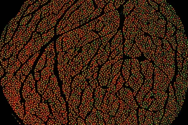
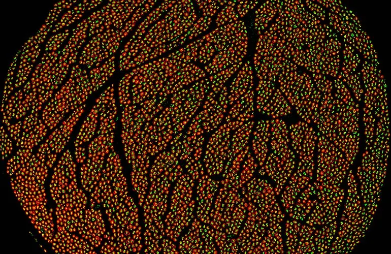
- Immunohistochemistry: Utilized specific fluorescent protein markers to illuminate individual nerve fibers, allowing scientists to differentiate between distinct fiber types and observe how they group together.
- Ultrasound Visualization: Captured real-time structural dynamics, bridging the gap between clinical imaging tools and microscopic realities.
Through this meticulous approach, the researchers generated spatial data detailing how fascicles branch, merge, and redistribute as the nerve travels downward from the cranium toward the thoracic and abdominal cavities.
Public Release and Open Science (2026)
Rather than keeping the trove of data proprietary, the Feinstein Institutes aligned with the open-science ethos of the NIH SPARC program. In mid-2026, the researchers uploaded their initial comprehensive dataset online, making high-resolution microCT scans, multi-level cross-sectional imagery, and detailed ultrasound videos freely available to global researchers, academics, and medical device manufacturers.
While this initial release represents a monumental baseline dataset, the research initiative is ongoing. The team expects to continuously upload supplemental datasets as their analysis deepens, culminating in comprehensive peer-reviewed publications once the broader longitudinal study reaches maturity.
Supporting Context & Metrics: The Anatomy and Promise of Vagus Neuromodulation
The Biological Superhighway
To understand the significance of the Feinstein Institutes’ mapping project, one must appreciate the sheer complexity of the vagus nerve (from the Latin vagus, meaning "wandering").
- Fiber Count: The nerve comprises two main trunks housing upwards of 200,000 distinct nerve fibers.
- Composition: It is a mixed nerve, meaning it contains both efferent fibers (carrying motor signals from the brain down to organs like the heart, lungs, and gut) and afferent fibers (carrying sensory information from those same organs back up to the brain, with roughly 80% of vagal fibers dedicated to sensory feedback).
- Systemic Reach: It touches nearly every major visceral organ, playing a direct role in heart rate variability, gastric motility, endocrine function, and immune system modulation.
From Discovery to Clinical Reality
The foundational work underpinning this mapping project traces back more than 30 years, when Dr. Kevin Tracey, President and CEO of the Feinstein Institutes, made a paradigm-shifting discovery: the nervous system directly regulates the immune system and controls inflammation.
Dr. Tracey’s research uncovered the "inflammatory reflex," a neural circuit in which the vagus nerve senses peripheral inflammation and signals the spleen and other organs to tone down the production of inflammatory cytokines. This foundational mechanism shattered the long-held dogma that the nervous and immune systems operate independently.

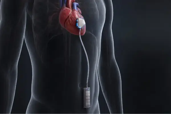
Dr. Tracey’s discoveries catalyzed a new therapeutic paradigm, leading directly to the invention and clinical translation of bioelectronic devices. Most notably, these insights birthed the SetPoint system, an FDA-approved vagus nerve stimulation device specifically engineered to treat severe rheumatoid arthritis by dampening systemic inflammation electronically. The device achieved a major clinical milestone when it was successfully implanted in its first commercial patients in August 2025.
However, designing devices like the SetPoint system historically relied on macroscopic approximations of nerve placement. With the newly completed anatomical map, engineers now possess the exact spatial coordinates of functional fibers, allowing for the creation of micro-electrodes that can selectively target anti-inflammatory pathways while avoiding fibers that control unwanted motor functions.
Official Statements from Key Thought Leaders
The completion of the human vagus nerve map has drawn widespread acclaim from the scientific community, highlighting its potential to rewrite the textbooks of human physiology and medical device engineering.
"This dataset represents a major step forward in bioelectronic medicine, offering the most detailed anatomical reconstruction of the human vagus nerve to date. For the first time, we can visualize the vagus nerve’s complex architecture that will allow us to design more precise, effective and safe neuromodulation therapies and devices."
— Dr. Stavros Zanos, Project Leader and Bioelectronic Medicine Researcher at the Feinstein Institutes
Dr. Zanos’s sentiment was strongly reinforced by institutional leadership, who emphasized the translational medical applications of decoding the body’s neural circuitry.
"Decoding the vagus nerve’s intricate language is an important advance for science and medicine. This knowledge will further empower researchers to re-engineer human biology and unlock novel therapies for future patients."
— Dr. Kevin Tracey, President and CEO of the Feinstein Institutes for Medical Research and pioneer of the inflammatory reflex
External evaluators and bioengineering consortiums have similarly lauded the project. By contributing this high-resolution atlas to the open-source SPARC ecosystem, the Feinstein team has effectively provided a foundational blueprint for global laboratories working on peripheral nerve interfaces, brain-computer interfaces (BCIs), and targeted organ stimulation.
Future Outlook: The Next Frontier in Bioelectronic Medicine
The completion of the human vagus nerve map marks not the end of a scientific journey, but the ignition of a new era in medicine. With the foundational architecture of the nerve now laid bare, several key developments are expected to reshape the medical device and therapeutic landscapes over the next decade.
1. Ultra-Precise Neuromodulation Devices
Current vagus nerve stimulation (VNS) systems—widely used for drug-resistant epilepsy, treatment-resistant depression, and recently, rheumatoid arthritis—wrap around the nerve trunk and deliver broad electrical fields. Armed with the Feinstein Institutes’ spatial map, next-generation device manufacturers can engineer multi-contact micro-cuff electrodes. These advanced implants will be capable of steering electrical current to specific fascicles, activating anti-inflammatory or cardioprotective fibers while leaving neighboring fibers untouched, thereby drastically reducing side effect profiles.
2. Expanding Indications for Chronic Disease
Chronic inflammation is recognized as a core driver of a vast spectrum of human pathologies, ranging from inflammatory bowel disease (IBD) and lupus to metabolic syndrome and cardiovascular disease. As bioelectronic devices become more anatomically precise, clinical trials are expected to expand rapidly into these previously intractable domains, offering drug-free alternatives for patients who fail conventional pharmaceutical regimens.
3. Accelerated Global Innovation via Open Science
Because the dataset is fully accessible via the SPARC portal, biomedical engineers, machine learning specialists, and neuroscientists worldwide can integrate this anatomical map into computational modeling software. Researchers can simulate electrical current spread through the nerve before ever entering a laboratory, drastically shortening the R&D cycle for medical hardware.
As ongoing datasets are uploaded and the scientific community continues to mine this unprecedented anatomical atlas, the vagus nerve will transition from a mysterious biological conduit into a fully deciphered highway of human health—ushering in an era where electricity cures disease with the surgical precision of a scalpel.