








Executive Overview
In a milestone achievement that bridges neuroscience, bioengineering, and clinical medicine, researchers at Northwell Health’s Feinstein Institutes for Medical Research have unveiled the world’s first comprehensive, high-resolution anatomical map of the human vagus nerve. Funded by a landmark $6.7 million grant from the National Institutes of Health (NIH), this three-year investigative effort marks a definitive paradigm shift in the burgeoning field of bioelectronic medicine.
For decades, the vagus nerve has been recognized as the principal superhighway of the autonomic nervous system—a bidirectional cable transmitting vital signals between the brainstem and virtually all major visceral organs. Yet, despite its critical role in regulating heart rate, respiratory rhythm, digestion, immune response, and inflammation, the precise internal topography of the nerve remained a biological black box. Medical device developers and neurosurgeons lacked a granular blueprint showing how individual nerve fibers and fascicles (bundles of fibers) are spatially organized and mapped to specific organ functions.
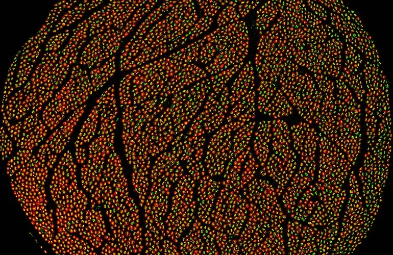
By analyzing 60 human vagus nerves from 30 donors using advanced micro-computed tomography (microCT), high-resolution ultrasound, and sophisticated immunohistochemistry with fluorescent protein markers, the Feinstein team has shattered this anatomical barrier. The resulting three-dimensional dataset provides an unprecedented look at the nerve’s microscopic architecture, revealing how individual fibers carry distinct physiological instructions.
By making this foundational data publicly accessible via the Stimulating Peripheral Activity to Relieve Conditions (SPARC) portal, the researchers have catalyzed a global movement. Bioengineers, device manufacturers, and clinical researchers can now leverage this atlas to design next-generation neuromodulation devices that are orders of magnitude safer, more precise, and more effective. This breakthrough promises to accelerate therapies for chronic, debilitating conditions ranging from rheumatoid arthritis and inflammatory bowel disease to drug-resistant epilepsy, heart failure, and severe psychiatric disorders.
Detailed Chronology: From Concept to Clinical Milestone
The realization of the world’s first human vagus nerve map is the culmination of years of targeted scientific inquiry, technological maturation, and meticulous multi-institutional collaboration.
The Foundational Breakthroughs (1990s–2020)
The scientific journey underpinning this achievement began over three decades ago, when Dr. Kevin Tracey, now President and CEO of the Feinstein Institutes for Medical Research, made a discovery that fundamentally altered modern immunology. Dr. Tracey and his team uncovered the inflammatory reflex—a neuro-immune pathway wherein the brain uses the vagus nerve to monitor and suppress systemic inflammation by signaling immune cells to halt the production of pro-inflammatory cytokines.
This foundational insight birthed the field of bioelectronic medicine, demonstrating that electrical impulses traveling along nerve pathways could replace or complement pharmaceutical drugs. Over the ensuing years, Dr. Tracey’s discoveries paved the way for clinical translation, culminating in the invention and eventual regulatory approval of vagus nerve stimulation (VNS) systems for inflammatory diseases, such as the SetPoint system for rheumatoid arthritis, which saw its first patient implants in August 2025.
However, as clinical applications expanded, a profound engineering bottleneck became apparent. Early VNS devices delivered electrical current broadly across the entire nerve trunk, frequently resulting in "off-target" side effects—such as voice alteration, coughing, bradycardia, or gastrointestinal discomfort—because the devices could not selectively stimulate only the fibers responsible for the targeted therapeutic effect.
Securing the NIH Investment (2021–2022)
Recognizing that the clinical potential of bioelectronic medicine was bottlenecked by a lack of precise anatomical knowledge, a research consortium led by the Feinstein Institutes pursued and secured a competitive $6.7 million grant from the National Institutes of Health (NIH). The objective was clear yet intensely challenging: systematically map the human vagus nerve from end to end, decoding its micro-architecture to establish a definitive reference standard for the scientific community.
The Three-Year Mapping Initiative (2023–2026)
Under the leadership of project director Dr. Stavros Zanos, the research team initiated a massive empirical study. Over a three-year period, the team procured and examined 60 vagus nerves sourced from 30 human cadaveric donors.
The investigative methodology required a multi-modal imaging approach capable of capturing both macroscopic trajectories and microscopic fiber-level resolution:
- MicroCT Imaging: Used to visualize the 3D trajectory of nerve bundles, branching patterns, and fascicular arrangements across the entire length of the nerve.
- High-Resolution Ultrasound: Deployed to capture dynamic structural variations and tissue density profiles.
- Immunohistochemistry & Fluorescent Protein Markers: Applied to tissue sections to stain specific neural structures, allowing researchers to differentiate motor fibers from sensory fibers and characterize the biochemical nature of individual pathways using advanced microscopy.
Public Release and Open-Science Sharing (Mid-2026)
Rather than keeping the high-value dataset proprietary, the Feinstein Institutes committed to an open-science framework, publishing the initial dataset online through the SPARC scientific data repository. This release included dynamic ultrasound videos, comprehensive microCT scans spanning the entire length of the nerve, and multi-level cross-sectional histological images. This immediate sharing empowers global research teams to accelerate their own device designs and computational models, fostering an era of collaborative bioelectronic innovation.
Supporting Context & Metrics: Decoding the Vagus Nerve
To understand the magnitude of the Feinstein Institutes’ achievement, one must examine the astonishing biological complexity of the vagus nerve itself.

Anatomical Scale and Composition
Derived from the Latin word vagus, meaning "wandering," the vagus nerve (cranial nerve X) is the longest and most complex cranial nerve in the human body. It emerges directly from the brainstem, travels down through the neck within the carotid sheath, passes through the thorax, and snakes its way into the abdomen, touching virtually every major visceral organ.
- Fiber Count: A typical human vagus nerve comprises over 200,000 individual nerve fibers split across two main bundles.
- Afferent vs. Efferent Signaling: Approximately 80% of vagus nerve fibers are afferent (sensory), carrying real-time status reports from peripheral organs—such as the gut, heart, and lungs—back to the central nervous system. Conversely, 20% are efferent (motor/secretory), carrying commands from the brain to modulate organ function.
- Myelination Spectrum: The nerve contains a heterogeneous mix of heavily myelinated A-fibers (fast-conduction motor and sensory signals), moderately myelinated B-fibers (preganglionic autonomic fibers), and unmyelinated C-fibers (slow-conduction visceral sensory and postganglionic autonomic fibers).
The Spatial Challenge in Neuromodulation
Before this comprehensive mapping initiative, neurosurgeons and biomedical engineers understood the macroscopic path of the vagus nerve (where it runs in the neck) and had general microscopic data regarding fiber types. However, what remained completely unknown was the spatial organization of fascicles—specifically, how fiber types are bundled together as the nerve travels down the body, and which specific fascicles innervate specific target organs.
Without this map, electrical stimulation was akin to broadcasting a radio signal across an entire city when you only wanted to reach a single house. By unlocking the precise topographic arrangement of fascicles, the Feinstein mapping data enables the creation of multi-contact, directional micro-electrodes. These advanced neural interfaces can steer electrical fields with pinpoint accuracy, stimulating only the specific fascicle associated with an anti-inflammatory response or glycemic control while sparing fibers that cause unwanted side effects.
Official Statements and Expert Perspectives
The release of the human vagus nerve atlas has drawn widespread praise from institutional leaders, neuroscientists, and bioelectronic medicine pioneers.
"This dataset represents a major step forward in bioelectronic medicine, offering the most detailed anatomical reconstruction of the human vagus nerve to date. For the first time, we can visualize the vagus nerve’s complex architecture that will allow us to design more precise, effective and safe neuromodulation therapies and devices."
— Dr. Stavros Zanos, Project Leader and Bioelectronic Researcher at the Feinstein Institutes
Dr. Zanos emphasizes that the transition from generalized nerve stimulation to fascicle-specific targeting will redefine clinical safety profiles. By understanding the exact geographic layout of the nerve, clinicians can tailor therapies to individual anatomical variations.
"Decoding the vagus nerve’s intricate language is an important advance for science and medicine. This knowledge will further empower researchers to re-engineer human biology and unlock novel therapies for future patients."
— Dr. Kevin Tracey, President and CEO of the Feinstein Institutes for Medical Research
Dr. Tracey, whose pioneering research decades ago linked neural signaling directly to immunological control, views this anatomical map as the ultimate realization of a long-term vision: treating chronic inflammatory diseases through software-controlled electrical impulses rather than lifelong chemical immunosuppression.
Future Outlook: The Next Frontier in Bioelectronic Medicine
The completion of this initial anatomical dataset is not an endpoint, but rather the foundation of an ongoing, expansive scientific endeavor. The Feinstein research team has confirmed that additional anatomical datasets and high-resolution imaging volumes will be continuously uploaded to public repositories as the study progresses toward its ultimate conclusions.
Transforming Medical Device Engineering
Armed with this high-resolution topographical map, medical device manufacturers are already pivoting toward advanced, next-generation neuromodulation architectures:
- Fascicular-Targeted Micro-Arrays: Engineers can now design cuff electrodes embedded with dozens of microscopic contact points, utilizing software algorithms to selectively activate individual fascicles based on real-time physiological feedback loops.
- Personalized Computational Modeling: Researchers are using the 3D datasets to build patient-specific digital twins of the vagus nerve, allowing surgeons to simulate electrical field dispersion prior to implanting a neurostimulator.
Expanding Clinical Indications
The clinical implications of precise vagus nerve mapping extend across a vast therapeutic landscape:
- Autoimmune and Inflammatory Disorders: Building upon the success of devices treating rheumatoid arthritis, precise stimulation protocols are currently being evaluated for Crohn’s disease, ulcerative colitis, and systemic lupus erythematosus.
- Metabolic and Cardiovascular Conditions: Targeted neuromodulation offers new hope for managing type 2 diabetes, obesity, and heart failure by modulating autonomic tone influencing glucose metabolism and cardiac output.
- Neurological and Psychiatric Illnesses: Enhanced selectivity promises to optimize treatments for drug-resistant epilepsy, major depressive disorder, post-traumatic stress disorder (PTSD), and migraine headaches while drastically reducing therapy-limiting side effects.
As the scientific community continues to mine this groundbreaking anatomical atlas, human biology and digital engineering converge ever closer. The Feinstein Institutes’ map of the vagus nerve has transformed a wandering, mysterious bundle of fibers into a clearly charted superhighway, ushering in a golden age of bioelectronic medicine where electrical impulses heal the body with unprecedented precision.