





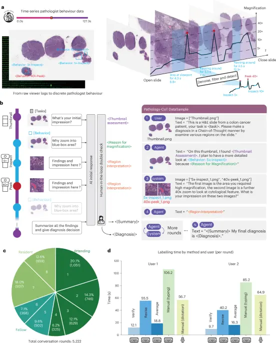
In a milestone study published in Nature Biomedical Engineering, a multi-institutional research team has unveiled Pathology-CoT, a pioneering framework designed to imbue vision-language models (VLMs) with the complex visual reasoning pathways characteristic of expert human pathologists. Spearheaded by corresponding author Zhi Huang of the Perelman School of Medicine at the University of Pennsylvania, alongside lead contributors such as Sheng Wang and R. Wu, the project bridges the long-standing gap between automated artificial intelligence (AI) triage and the nuanced, dynamic navigation required to interpret whole-slide images (WSIs).
Diagnostic pathology has historically relied on the meticulous analysis of digital WSIs—gigapixel-scale panoramas where a single misstep can mean the difference between a benign finding and an aggressive malignancy. While prior computational systems have attempted to automate slide reading using static computer vision models, they often lack the step-by-step cognitive transparency—the "chain of thought"—that human specialists employ. Pathologists do not merely stare at a slide; they pan, zoom, reassess normal tissue architecture, and hunt for subtle irregularities through an iterative process of structural hypothesis and verification.
Pathology-CoT mimics this exact behavioral methodology. By capturing, cleaning, and feeding expert navigation trajectories into advanced VLMs, the framework trains artificial agents to inspect regions of interest (ROIs) with the precision, stability, and diagnostic intuition of seasoned clinicians. This comprehensive report explores the architecture of the Pathology-CoT pipeline, its performance across complex clinical cohorts such as colorectal cancer lymph node metastasis and dermatopathology, and the broader implications for computational pathology.
Detailed Chronology & Methodology: Capturing and Translating Expert Behavior
The development of Pathology-CoT required surmounting a fundamental hurdle in computational biology: translating the fluid, highly variable physical interactions of a pathologist navigating a digital microscope into a structured, machine-readable format.
1. The AI Session Recorder and Behavioral Preprocessing
To capture how experts actually look at WSIs, the researchers implemented a robust behavioral capture methodology. When pathologists examine slides, their movements generate noisy, high-frequency viewport data—frequent overlapping pans, erratic zoom adjustments, and variable dwell times.
To transform this raw stream into actionable training data, the team built an AI Session Recorder pipeline, validated against biological eye-tracking data. The preprocessing pipeline operates across six distinct stages:
- Raw Behaviour Ingestion: Capturing initial, noisy viewport coordinates and mouse movements.
- Initial Action Segmentation: Grouping raw events into discrete behavioral classes using temporal heuristics, specifically separating StayInspect (focused observation periods) from PanInspect (transitional movement across the slide).
- Filtering Overview Actions: Stripping away very large, low-magnification bounding boxes that correspond merely to non-diagnostic, macro-level slide overviews.
- Merging Overlapping Actions: Combining spatially proximal actions with high Intersection over Union (IoU) scores into single, consolidated inspection zones.
- Pruning Containing Actions: Removing larger, redundant actions that fully encapsulate smaller, highly specific inspection targets to eliminate noise.
- Normalizing Action Bounding Boxes: Resizing the final set of actions to standard dimensions corresponding to discrete magnification levels (e.g., $5times$, $10times$), thus establishing a consistent, clean input format for the downstream vision-language model.
Validation tests demonstrated striking consistency. When comparing session recordings from the same attending pathologist across different trials, the AI Session Recorder yielded remarkably stable and reproducible ROI results, proving its reliability as an objective behavioral metric.
2. Training the Visual Chain-of-Thought Agents
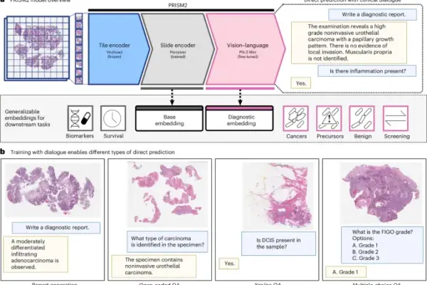
With a clean dataset of navigational actions, the team trained specialized behavior predictors and vision-language agents (such as the advanced Pathology-o3 architecture). Unlike standard multi-modal models that are fed a static image and prompted for an instantaneous diagnosis, Pathology-CoT agents are trained on the temporal sequence of visual attention.
The models learn how to zoom into problematic zones, when to step back for structural anatomical context, and how to deliberate over morphological anomalies—mirroring the cognitive loop of a clinical diagnostic session.
Supporting Context & Metrics: Performance Stratification and Validation
To rigorously evaluate Pathology-CoT, the team deployed the framework across two distinct, highly challenging clinical datasets: the Stanford colorectal cancer (CRC) lymph node cohort and the DROID skin cancer cohort. The results highlight substantial performance gains over conventional proprietary and open-source models.
Colorectal Cancer Lymph Node Metastasis
Detecting micrometastases and isolated tumor cells (ITCs) in lymph nodes is notoriously difficult due to the scarcity of tumor cells and the absence of clear desmoplastic reactions. The researchers stratified performance across three clinical difficulty tiers:
- Isolated Tumor Cells (ITCs, $le 0.2text mm$, $n=5$): The ultimate test of fine-grained detection.
- Micrometastases ($0.2text–2text mm$, $n=13$): Intermediate burden cases requiring precise spatial targeting.
- Macrometastases ($>2text mm$, $n=29$): Larger, more overt tumor formations.
In comparative benchmarks, Pathology-o3 outperformed the best proprietary models (such as GPT-4.1 and GPT-5.2 variants), top open-source baselines (InternVL3), and legacy architectures (Llama-4). Visualizations of zoom-in behaviors revealed that while general-purpose models often became disoriented or misallocated attention, Pathology-o3 mirrored the precise navigation patterns of attending pathologists—successfully homing in on elusive tumor clusters lacking desmoplastic reactions.
Quantitative Attribution of Attention Drivers in Dermatopathology
Using the DROID segmentation ontology, the researchers conducted an ablation and attribution analysis on 960 highest-confidence ROIs predicted by the YOLOv8-Large behavior predictor model.
- Screening-Like Behavior: Interestingly, the analysis revealed that the majority of model attention (62.9%) was allocated to normal anatomical structures (e.g., healthy epidermis, skin appendages) rather than explicit pathology. This reflects a natural "screening-like" pattern, where human experts first establish baseline normal architecture before zoning in on abnormalities.
- Pathological Subdivisions: Of the ROIs classified explicitly as "Pathology" ($N=340$), the model successfully identified both frank neoplasms (basal cell carcinoma, melanocytic neoplasms, squamous cell carcinoma) and subtle non-neoplastic findings (33.5% of pathological ROIs), which included inflammatory responses, fibrosis, and cellular infiltration.
- Model Scale Ablation: Testing YOLOv8 architectures of increasing capacity (Nano to Large) demonstrated that smaller models were frequently misled by local artifacts (mistaking slide ink or blood pools for ROIs), whereas larger models demonstrated a sophisticated understanding of global tissue architecture, safely ignoring local distractors.
Official Statements & Data Availability
The research was conducted under strict ethical standards, with the authors declaring no competing interests. The project received foundational support via startup funding from the Perelman School of Medicine at the University of Pennsylvania (awarded to Z. Huang), which played no role in study design, data collection, or manuscript preparation. Peer review oversight was provided by Nature Biomedical Engineering, alongside external reviewers including Narayan Hegde and Hamid R. Tizhoosh.
To ensure total transparency, reproducibility, and collaborative growth within the computational biology community, the study’s core assets have been made fully accessible:
- Source Code & Datasets: The Pathology-CoT Chain-of-Thought dataset for the skin cancer cohort, alongside trained behavior predictors for both the Stanford CRC lymph node and skin cancer cohorts, is publicly available on GitHub at github.com/zhihuanglab/Pathology-CoT.
- AIDA Data Hub Integration: Whole-slide images and expert annotations from the LNCO2 dataset and the DROID skin dataset are accessible via the AIDA Data Hub (
datahub.aida.scilifelab.se) upon formal application and institutional approval.
Future Outlook
The introduction of Pathology-CoT marks a paradigm shift in how artificial intelligence interacts with medical imagery. By moving away from "black-box" classification—where an algorithm outputs a binary diagnosis without explanation—toward behavioral emulation, the framework provides a verifiable audit trail of AI reasoning.
Future developments will likely focus on scaling Pathology-CoT across broader oncological subspecialties, integrating multi-omic data streams into the visual reasoning loop, and deploying real-time AI session recorders in clinical training environments. For resident pathologists, these tools could serve as interactive tutors, analyzing navigation habits and providing real-time feedback on diagnostic blind spots. As computational pathology continues its rapid evolution, frameworks like Pathology-CoT ensure that artificial intelligence remains not merely a computational tool, but a transparent, trustworthy cognitive partner in clinical medicine.