





Executive Overview
In a monumental leap forward for precision medicine and neurodegenerative research, scientists at Johns Hopkins Medicine have unveiled a groundbreaking application for laboratory-grown human brain tissue. By cultivating pea-sized clusters of brain cells derived directly from the blood samples of Alzheimer’s disease patients, researchers have successfully demonstrated that these miniature models—known as organoids—can predict individual patient responses to psychiatric medications.
Alzheimer’s disease, the most common form of dementia, currently affects more than 7 million Americans. While cognitive decline remains the hallmark of the condition, nearly all patients suffer from severe neuropsychiatric symptoms, including debilitating anxiety, profound depression, and intense agitation. To manage these symptoms, clinicians frequently prescribe selective serotonin reuptake inhibitors (SSRIs). However, because the underlying molecular pathology of Alzheimer’s varies wildly from person to person, the clinical efficacy of these drugs is notoriously unpredictable. Some patients experience meaningful relief, while others see no benefit at all, suffering only from side effects.
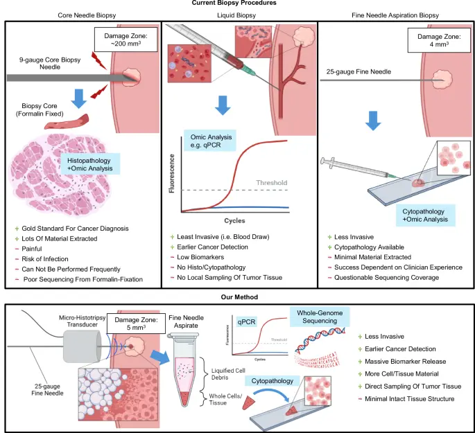
The new Johns Hopkins study, published in Alzheimer’s & Dementia: The Journal of the Alzheimer’s Association, bridges this critical clinical gap. By creating hundreds of patient-specific hindbrain organoids, the research team not only modeled the complex cellular environment of the human brain but also tracked how these tissues responded molecularly to escitalopram oxalate, a widely used SSRI. Furthermore, the team discovered that these organoids secrete extracellular vesicles—microscopic biological cargo containers—which mirror the molecular state of the tissue and could eventually serve as revolutionary diagnostic "liquid biopsies."
Supported in part by funding from the National Institutes of Health (NIH), the Paul G. Allen Frontiers Foundation, and the Richman Family Precision Medicine Center of Excellence in Alzheimer’s Disease, this expansive study marks a pivotal shift away from generalized trial-and-error prescribing. Instead, it ushers in an era where lab-grown neural avatars could dictate tailored psychiatric interventions for vulnerable dementia patients.
Detailed Chronology: From Blood Cells to Predictive Brain Models
The methodology behind this breakthrough marries cutting-edge stem cell biology with high-throughput molecular screening. To understand how Johns Hopkins researchers achieved this feat, it is necessary to examine the step-by-step evolution of the project, from clinical recruitment to laboratory cultivation and drug testing.
Phase 1: Patient Recruitment and Cellular Reprogramming
The foundation of the study was built upon human tissue samples provided with informed consent by individuals living with Alzheimer’s disease, alongside healthy control participants, through the NIH-funded Johns Hopkins Alzheimer’s Disease Research Center.
Rather than requiring invasive brain biopsies, researchers extracted routine blood samples. Through cellular reprogramming, the team returned these mature blood cells to a primitive, pluripotent stem cell-like state. These induced pluripotent stem cells (iPSCs) possess a remarkable biological superpower: they retain the exact genetic and molecular blueprint of the donor while holding the capacity to develop into any cell type in the human body.
Phase 2: Cultivating the Hindbrain Organoids
With the iPSCs established, the researchers directed the cells to differentiate specifically into neural lineages, focusing particularly on the hindbrain. The hindbrain is an evolutionarily ancient region located at the base of the skull, responsible for regulating vital autonomic functions such as respiration, heart rate, and sleep cycles. Within this region, the team specifically cultivated specialized neurons capable of producing and responding to serotonin, the primary neurotransmitter targeted by standard antidepressants.
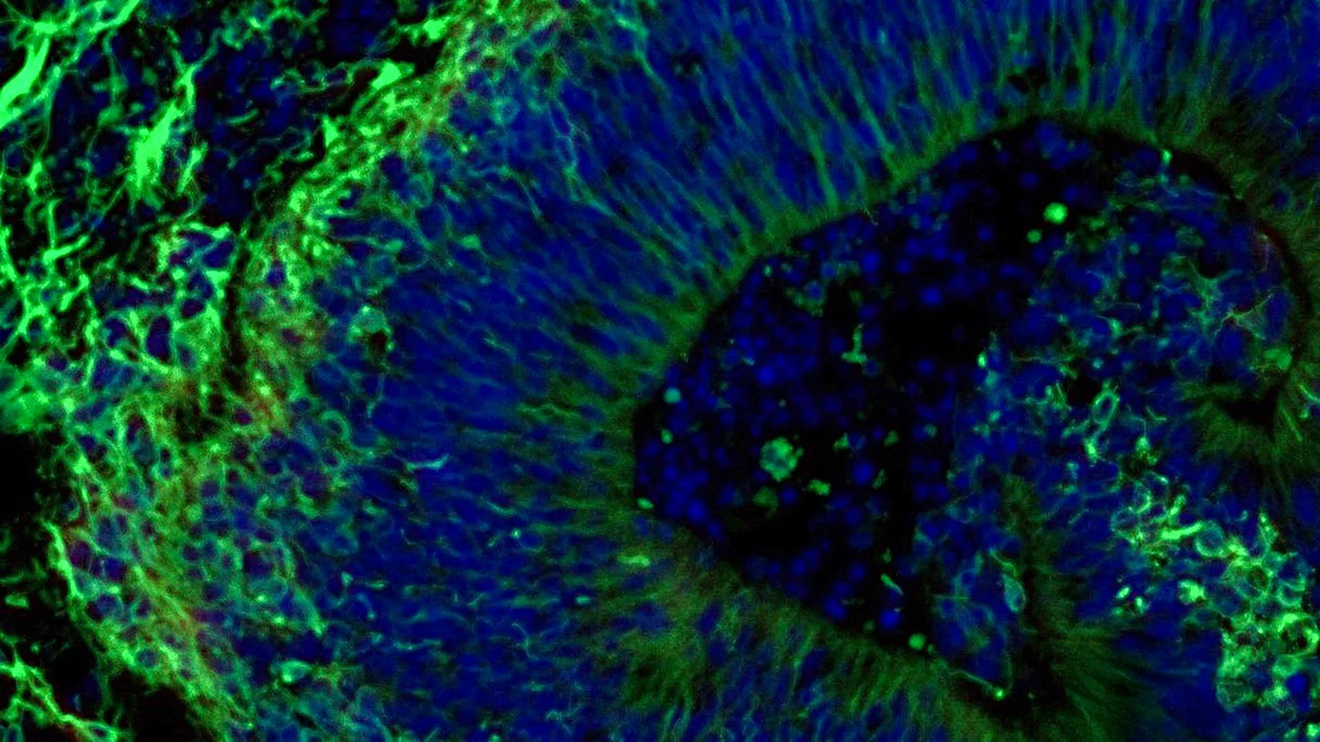
Through meticulous chemical signaling and three-dimensional suspension culture, the cells naturally organized themselves into pea-sized aggregates. This study stands out as one of the largest brain organoid investigations ever executed in the field of dementia research, encompassing hundreds of distinct organoids representing both Alzheimer’s patients and healthy controls.
Phase 3: Molecular Profiling and Drug Administration
Once mature, the organoids were subjected to deep molecular profiling. Researchers compared the protein expression profiles of the Alzheimer’s-derived organoids against those from healthy individuals. The results were striking: the patient-derived organoids faithfully replicated core molecular signatures of Alzheimer’s disease, displaying clear aberrations in proteins responsible for intercellular communication, neuroinflammation, and synaptic function.
The team then exposed the organoids to escitalopram oxalate. Following treatment, the researchers analyzed how the molecular machinery within the tissue reacted. In a subset of the Alzheimer’s-derived organoids, the antidepressant successfully upregulated proteins involved in serotonin signaling and synaptic communication—the exact pathways intended to be altered by the medication. Crucially, however, other patient organoids exhibited little to no molecular response, mirroring the disparate outcomes observed in real-world clinical psychiatry.
Supporting Context & Metrics: Unlocking the Power of Extracellular Vesicles
Beyond the cellular responses observed within the organoid tissue itself, the Johns Hopkins team made a secondary discovery that could fundamentally change how Alzheimer’s is diagnosed, staged, and monitored over time.
The Secretome and Liquid Biopsies
Cells constantly communicate by shedding microscopic membrane-bound packets called extracellular vesicles (EVs). These tiny biological parcels carry proteins, lipids, and nucleic acids from their parent cells into surrounding biofluids. The researchers collected and analyzed the extracellular vesicles secreted by the hindbrain organoids both before and after escitalopram treatment.
Biochemical analysis revealed that these EVs were packed with proteins essential for normal neurological function, such as neural signaling, memory consolidation, and neurotransmitter release. However, significant deficits were identified in the vesicles derived from Alzheimer’s organoids compared to healthy controls. Specifically, levels of key synaptic proteins—including RAB3A, NSF, and ATCAY—were markedly suppressed in the Alzheimer’s models, highlighting disrupted communication channels between brain cells.
Following the administration of escitalopram, the cargo inside the EVs changed dynamically. Vesicles from responsive organoids showed notable increases in proteins linked to serotonin pathways, whereas non-responsive organoids remained static.
Quantitative Impact and Scope
- 7 Million+: The number of Americans currently living with Alzheimer’s disease, nearly all of whom will experience neuropsychiatric symptoms like depression, anxiety, and agitation.
- Hundreds: The unprecedented volume of individual brain organoids analyzed in this study, making it one of the largest datasets of its kind.
- Key Proteins Identified: Significant baseline reductions in RAB3A, NSF, and ATCAY proteins within Alzheimer’s models, providing robust, quantifiable biomarkers of disease progression.
- Multi-Agency Support: Backed by critical grants from the NIH (including awards T32 AG058527, R01AG052510, P30AG066507, and multiple RF1/R01 mechanisms), alongside the Paul G. Allen Frontiers Foundation and the Richman Family Precision Medicine Center.
Official Statements and Expert Perspectives
The implications of this research extend far beyond academic pathology, offering a beacon of hope for clinicians struggling to manage the harrowing psychiatric symptoms of dementia.
Study leader Vasiliki Machairaki, Ph.D., associate professor of genetic medicine at the Johns Hopkins University School of Medicine, emphasized the transformative potential of patient-derived tissue models during an interview discussing the findings:
"Our study suggests that large-scale, patient-derived brain organoids and the vesicles they secrete can help us stage Alzheimer’s disease, investigate the mechanisms that drive it, and assess how patient subgroups may respond to different treatments."
Addressing the clinical challenge of prescribing antidepressants to dementia patients—where physicians are often forced into a cycle of trial-and-error—Machairaki elaborated on the utility of the screening platform:
"We used these organoids to model how some patients’ tissue may respond to a commonly prescribed SSRI. On a large-scale level, our model may eventually be used to identify subgroups of patients, based on underlying molecular mechanisms, who are more likely to respond to certain drugs and thus help us to create precise, targeted treatments in the long run."
The multidisciplinary research team behind this publication represents a powerhouse of academic and industrial collaboration. Alongside Dr. Machairaki, the study’s co-authors include Rachel Boyd, Daiyun Dong, Ram Sagar, Waqar Ahmed, Xenia Androni, Paul Rosenberg, Constantine Lyketsos, and Kenneth Witwer from Johns Hopkins University; Anton Iliuk from Tymora Analytical Operations; and Anton Porsteinsson from the University of Rochester School of Medicine and Dentistry. In accordance with university policies, the authors declared no related conflicts of interest.
Future Outlook: Toward Vascularized Organoids and Liquid Biopsies
While the current findings represent a monumental step forward, the Johns Hopkins team views this study as merely the foundation for a much grander translational vision.
Engineering More Complex Human Tissues
Real human brain tissue is vastly more complex than the static organoids utilized in the initial phases of this study. To bridge the gap between bench science and clinical reality, Dr. Machairaki and her colleagues are already working on developing next-generation organoids. Future iterations will incorporate functional immune cells (such as microglia, which drive the neuroinflammation characteristic of Alzheimer’s) and vascular-like networks designed to mimic the brain’s blood supply. By introducing these biological systems, the tissue models will more accurately replicate the microenvironment of a living human brain.
The Ultimate Goal: A Non-Invasive Diagnostic Tool
The most exciting long-term prospect of this research lies in the clinical translation of extracellular vesicles. If researchers can validate that the EV protein signatures discovered in lab-grown organoids match those found circulating in the blood or cerebrospinal fluid of living patients, it could unlock the holy grail of neurodegenerative diagnostics: a reliable, blood-based "liquid biopsy."
Such a test could fundamentally transform clinical care by:
- Diagnosing Alzheimer’s Disease earlier and with greater molecular precision than current cognitive tests allow.
- Staging the progression of neurodegeneration accurately over time.
- Identifying distinct disease subtypes, allowing physicians to match a patient’s unique molecular profile to the pharmaceutical intervention most likely to provide relief.
Although these advanced clinical applications remain on the horizon, the Johns Hopkins study establishes an undeniable proof-of-concept. By translating a patient’s blood cells into living, predictive neural tissue, science has moved one step closer to decoding the complexities of Alzheimer’s disease and delivering genuinely personalized medicine to millions in need.