









Executive Overview
Bladder cancer remains one of the most stubborn, pervasive, and clinically challenging malignancies in modern oncology. Characterized by high rates of post-treatment recurrence—affecting up to 80% of patients—the disease frequently forces physicians and patients into a difficult corner. Current standard-of-care interventions often demand aggressive repeat surgeries or, in severe cases, radical cystectomy, the complete surgical removal of the bladder. While life-saving, total bladder removal dramatically alters a patient’s quality of life, leaving them permanently incontinent and requiring lifelong adjustments.
Now, a pioneering multidisciplinary research team led by the University of Massachusetts Amherst (UMass Amherst) is setting out to change this paradigm. Funded by a substantial five-year grant of up to $3.45 million from the National Institutes of Health (NIH), the team is repurposing and scaling an advanced soft-tissue ablation technique. Their goal is remarkably ambitious yet elegantly simple: to eliminate hidden, pre-cancerous, and malignant cells across the entire inner lining of the bladder without harming the organ itself or requiring its surgical removal.
Led by principal investigator Govind Srimathveeravalli, the research endeavor adapts irreversible electroporation (IRE)—a non-thermal tissue ablation method clinically proven in cardiac and oncological applications—into a flexible, catheter-delivered therapy. By targeting the delicate, three-to-four-cell-thick inner layer of the bladder known as the urothelium, the UMass Amherst team hopes to systematically "exfoliate" diseased cellular material, preempting cancer emergence and allowing healthy surrounding tissue to regenerate naturally. Utilizing cutting-edge "bladder-on-a-chip" microphysiological models and advanced computer simulations, this translational project bridges the gap between bioengineering and clinical oncology, offering a beacon of hope to hundreds of thousands of patients worldwide.
Detailed Chronology of the Research Initiative
The journey toward developing this novel ablation technology is rooted in years of iterative bioengineering breakthroughs, culminating in the recent major influx of federal backing and advanced laboratory modeling.
The Genesis of a Novel Approach
For decades, the medical device and oncology communities have grappled with the unique anatomical and physiological challenges posed by the human bladder. Unlike solid tumors located in the liver, prostate, or kidneys—which present as localized, discrete masses—bladder cancer frequently manifests as a diffuse field of genetic mutations spread across an expansive, highly elastic, and irregularly shaped mucosal surface.
Dr. Govind Srimathveeravalli and his colleagues at UMass Amherst began investigating how existing surgical and ablation tools could be modified to address these diffuse fields. Traditional surgical resections, known as transurethral resection of bladder tumor (TURBT), often miss microscopic pockets of diseased tissue hiding within the folds of the bladder wall. This invisibility cloak of hidden malignancy is the primary driver behind the staggering 80% recurrence rate.
Recognizing that localized needle electrodes were insufficient for treating an entire organ lining, the UMass team conceptualized a fundamental shift in strategy. Instead of hunting for individual tumors after they manifest, why not treat the entire vulnerable terrain proactively? This conceptual turning point led them directly to irreversible electroporation.
Securing the $3.45 Million NIH Milestone
As the foundational engineering concepts matured, the team sought the rigorous validation and capital required to push the technology from academic benchtop prototypes toward human clinical trials. That validation arrived when the National Institutes of Health awarded the team a five-year grant totaling up to $3.45 million.
This federal funding serves as the financial and structural backbone for the project’s next phase. It enables the UMass Amherst researchers to scale up their laboratory operations, expand their collaborative network, build sophisticated testing models, and systematically de-risk the technology across pre-clinical milestones. The grant reflects not only the high-risk, high-reward nature of the proposal but also the pressing, unmet clinical need for bladder-sparing therapies.

Integration of Advanced Microphysiology Systems
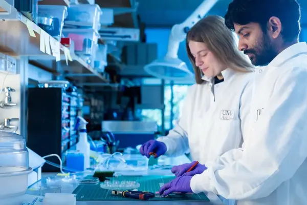
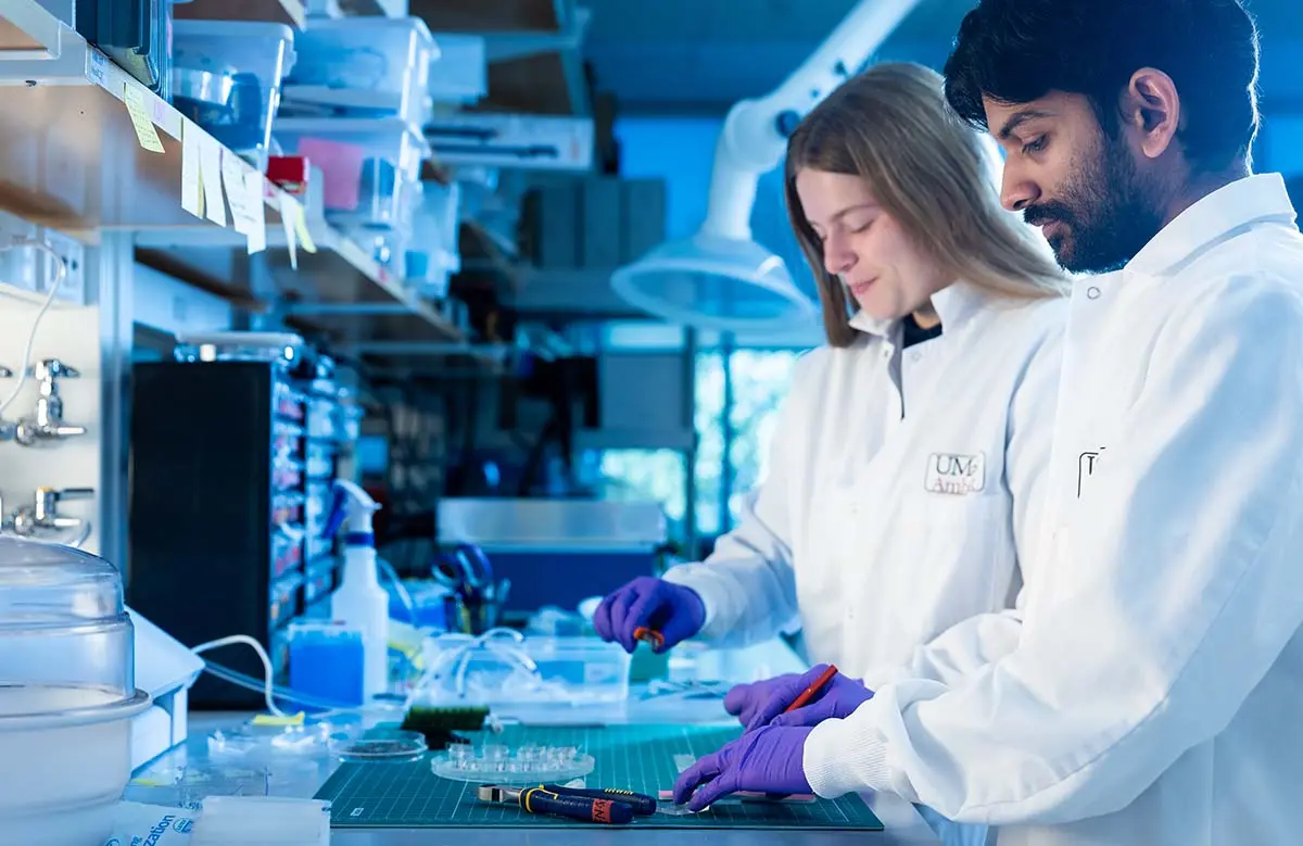
With funding secured, the research team implemented a modern testing infrastructure that relies heavily on alternative research methods, bypassing the limitations of traditional animal testing models. Collaborating across disciplines, the team is manufacturing "bladders on chips"—microphysiological systems populated with patient-derived cells.
These chips replicate the micro-architecture, mechanical stretch, and fluid dynamics of the human urinary bladder in a laboratory dish. By combining these biological models with robust computer simulations, the researchers can map out the precise electrical field parameters required to achieve uniform cell removal. This methodology ensures that their findings are deeply translational, highly reproducible, and rigorously optimized before any first-in-human clinical evaluations begin.
Supporting Context & Metrics: The Science of Irreversible Electroporation (IRE)
To truly grasp the significance of the UMass Amherst project, one must examine the underlying biophysics of irreversible electroporation and the stark clinical realities of bladder cancer epidemiology.
Understanding Irreversible Electroporation (IRE)
Irreversible electroporation is a revolutionary tissue ablation modality that operates on a fundamentally different principle than traditional thermal ablation techniques like radiofrequency ablation (RFA), cryoablation, or laser therapy.
- Non-Thermal Mechanism: Traditional ablation relies on extreme heat or cold to coagulate or freeze proteins, destroying cells indiscriminately. This thermal energy often damages adjacent structural tissues, nerves, and blood vessels, posing significant clinical risks when applied to delicate organs.
- Electrical Disruption: IRE, by contrast, delivers ultra-short, high-voltage electrical pulses via specialized electrodes. These pulses create microscopic nanoscale defects—known as pores—within the lipid bilayer of cell membranes.
- Cellular Necrosis: When the electrical parameters exceed a specific threshold, the cellular membrane damage becomes permanent (irreversible). The cell loses its homeostasis, leading to programmed cell death (apoptosis) and tumor necrosis.
- Preservation of Extracellular Matrix: Because IRE targets the lipid structure of cell membranes rather than relying on thermal destruction, the underlying extracellular matrix, collagen scaffolding, blood vessels, and ductal structures remain intact. This preservation is critical for promoting rapid, healthy tissue regeneration.
The PFA Connection in Modern Medicine
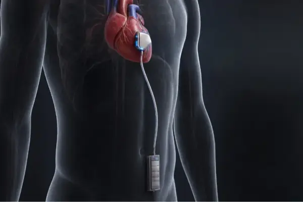
The biophysical principles underpinning IRE are not entirely unfamiliar to modern medicine. In recent years, pulsed-field ablation (PFA)—a direct technological cousin of IRE—has taken the electrophysiology and cardiology sectors by storm. PFA catheters are now widely utilized to treat cardiac arrhythmias, specifically atrial fibrillation (AFib), by selectively ablating aberrant heart tissue cells with microsecond electrical pulses while leaving surrounding vascular and neural structures unharmed.
By leveraging the lessons learned from PFA device design and manufacturing—particularly regarding flexible catheter delivery systems and precise energy generators—the UMass Amherst team is positioned to accelerate the translation of these concepts from cardiology into urological oncology.
Anatomy of the Urothelium
The primary anatomical target of this new technology is the urothelium, a specialized layer of stratified epithelial cells that lines the entire inner surface of the urinary tract, including the renal pelvis, ureters, bladder, and proximal urethra.
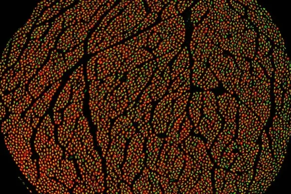
In the bladder, the urothelium acts as an impermeable barrier against toxic waste products present in urine. However, this dynamic cell layer is approximately 0.1 millimeters thick—roughly three to four cells deep. Because it is constantly exposed to chemical carcinogens filtered by the kidneys, it is a prime site for oncogenic mutations.
The breakthrough insight driving the UMass project is the realization that the urothelium possesses a remarkable capacity for natural regeneration. By designing a catheter capable of applying precise, uniform IRE pulses across the entire inner surface of the bladder, clinicians could theoretically "exfoliate" the diseased urothelial lining entirely. The damaged, pre-malignant cells are cleared away, and healthy stem cells within the underlying tissue regenerate a pristine, cancer-free urothelium.

The Clinical Burden of Bladder Cancer
The necessity for such an innovation is underscored by compelling epidemiological metrics:
- High Incidence: Bladder cancer is consistently ranked among the most common malignancies globally, accounting for hundreds of thousands of new diagnoses annually.
- Recurrence Rates: Up to 80% of superficial or non-muscle-invasive bladder cancer patients experience tumor recurrence following initial surgical removal, necessitating lifelong monitoring and repeated interventions.
- The Cost of Care: Bladder cancer carries one of the highest cumulative lifetime per-patient treatment costs of any cancer, driven largely by the frequency of surveillance cystoscopies and recurrent surgical resections.
- Surgical Morbidity: When recurrence escalates or progresses to muscle invasion, radical cystectomy becomes unavoidable. This major open or robotic surgery involves urinary diversion (such as an ileal conduit or neobladder creation), fundamentally altering patient anatomy and impacting long-term physical and psychological well-being.
Official Statements & Expert Perspectives
The research initiative represents a convergence of engineering brilliance and clinical pragmatism, driven by leaders who recognize both the promise of the technology and the hurdles of clinical translation.
In an official university news release detailing the project, lead researcher Govind Srimathveeravalli articulated the core frustration that inspired the innovation:
"The patient’s bladder has these diseased cells that we don’t know what they look like, we don’t know where they’re hiding, and we can’t get rid of them."
Traditional diagnostics—ranging from white-light cystoscopy to advanced imaging—frequently fail to resolve microscopic fields of cellular atypia scattered across the vast expanse of the bladder mucosa. Srimathveeravalli emphasized that fighting an enemy you cannot see requires shifting the strategic objective from targeted tumor destruction to comprehensive field management:
"Why allow the progression of diseased urothelial cells that form the inner lining of the bladder to malignancy? Why not exfoliate this cell layer where the diseased cells are hiding, thereby preempt the emergence of cancer and allow the healthy cells in the layer to regenerate?"
Addressing the technical challenges of scaling IRE from a focal needle electrode to a comprehensive, organ-wide catheter treatment, Srimathveeravalli outlined the vision for the UMass-led team:
"We want to transform IRE technology from a single-area, ultra-targeted treatment into a flexible, catheter-based treatment to target the three-to-four cells thick innermost layer of the entire bladder wall."
Looking toward the horizon, Srimathveeravalli expressed strong confidence in the development pipeline, particularly regarding the integration of physiological modeling to de-risk human application:

"If we are successful in showing that this is safe and effective, it is very well positioned to take this into clinical trials as the next step."
Future Outlook: Toward Clinical Translation and Paradigm Shift
As the UMass Amherst research team embarks on this five-year NIH-backed endeavor, the medical technology and oncology communities are watching closely. The implications of a successful bladder-sparing ablation device extend far beyond the urology suite.
Overcoming Engineering and Clinical Hurdles
While the theoretical framework is sound, significant engineering challenges remain. Scaling an electrical field to conform seamlessly to the complex, changing geometry of an expanding and contracting human bladder requires sophisticated energy delivery algorithms. Researchers must precisely calibrate voltage, pulse duration, and electrode configuration to ensure complete eradication of the 0.1-millimeter urothelial layer without penetrating too deeply into the muscularis propria—the muscular wall of the bladder responsible for organ contraction and urination.
The utilization of patient-derived "bladders on chips" provides a vital testing ground to solve these precise volumetric and electrical challenges. By simulating thousands of treatment variations computationally and validating them against microphysiological tissue models, the UMass team can fine-tune the delivery catheter’s geometry before advancing to animal models and, ultimately, human trials.
Transforming Patient Outcomes
If successful, this technology threatens to upend decades of entrenched clinical practice. A catheter-delivered, non-thermal ablation procedure performed under conscious sedation or light anesthesia could replace repeated surgical resections and indefinitely postpone—or entirely prevent—the need for radical cystectomy.
Patients would retain their native bladders, avoid the psychological and physical trauma of major reconstructive surgery, maintain urinary continence, and enjoy a vastly superior quality of life. Furthermore, by preempting the progression of dysplastic urothelial cells into invasive carcinoma, the healthcare system could experience dramatic reductions in long-term treatment costs and mortality rates associated with advanced metastatic bladder cancer.
A New Era for Electroporation in Oncology
Beyond urology, the success of UMass Amherst’s organ-lining exfoliation technique could establish a powerful new blueprint for treating epithelial cancers throughout the human body. Mucosal surfaces in the gastrointestinal tract, bronchial tree, and gynecological tracts often present similar challenges regarding diffuse field cancerization and high recurrence rates.
By proving that irreversible electroporation can be scaled safely and effectively to treat entire continuous tissue layers, Srimathveeravalli and his team are not only rewriting the future of bladder cancer care—they are opening an entirely new chapter in bioelectronic medicine.