











Executive Overview
Artificial intelligence has entered a transformative era in digital healthcare, promising to shift medicine from reactive treatment to proactive, precision-based therapies. Nowhere is this promise more pronounced—or the technological hurdles more formidable—than in the field of computational pathology. As high-resolution digital whole-slide images (WSIs) replace traditional glass microscope slides, the medical community stands on the precipice of a diagnostic revolution. However, realizing this potential requires more than just high-powered algorithms; it demands robust artificial intelligence foundation models capable of generalizing seamlessly across diverse datasets, varying tissue types, and complex clinical tasks.
Until recently, the comparative performance and true generalizability of these advanced foundation models remained incompletely characterized, creating a barrier to clinical adoption. A monumental new benchmark study published in Nature Communications (2026), titled "A benchmark study of vision and pathology foundation models for computational pathology," offers a definitive look at the current landscape. Led by corresponding author Olivier Gevaert and his research team at Stanford University, alongside contributions from Rohan Bareja, Fernando Carrillo-Perez, and Y. Zheng, the study delivers an exhaustive evaluation of 32 distinct AI foundation models divided into four major categories.
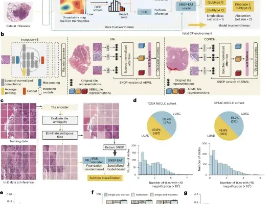
Evaluating these systems across slide-level and patch-level tasks utilizing gold-standard repositories such as The Cancer Genome Atlas (TCGA), the Clinical Proteomic Tumor Analysis Consortium (CPTAC), and various external and out-of-domain datasets, the research sheds light on what works, what falls short, and how the computational pathology community must move forward. The study’s primary takeaway is both encouraging and cautionary: while pathology-specific vision models consistently lead the pack, differences among top performers are often negligible and task-dependent, model scale does not reliably predict success, and strategic ensembling can unlock unprecedented diagnostic power. To foster ongoing innovation and transparency, the research team has launched PathBench, an open-access platform available at https://pathbench.stanford.edu/, designed to serve as a continuous evaluation standard for the scientific community.
Detailed Chronology and Methodological Architecture
The genesis of this comprehensive benchmarking effort lies in a critical bottleneck within modern digital pathology. Over the past decade, hundreds of machine learning models have been trained on gigapixel-sized pathology images. Yet, researchers and clinical practitioners have lacked a standardized, unbiased framework to compare them directly. Different models are often evaluated on disparate internal datasets using varied metrics, making it nearly impossible for hospitals, research institutions, and regulatory bodies to determine which architecture is best suited for clinical deployment.
To address this information gap, the Stanford-led research consortium embarked on a systematic evaluation timeline, establishing a rigorous taxonomy to classify and test 32 state-of-the-art foundation models. The models were categorized into four distinct architectural groups:
- General Vision Models (VM): Broad computer vision architectures originally trained on natural images (such as standard ImageNet-based or self-supervised transformers) that are subsequently adapted for medical image analysis.
- General Vision-Language Models (VLM): Multimodal systems trained on both vast repositories of images and natural language text, allowing them to process visual features alongside clinical reports or textual prompts.
- Pathology-Specific Vision Models (Path-VM): Models explicitly pretrained from the ground up on massive, curated corpuses of histopathology whole-slide images, enabling them to capture cellular structures, staining variations, and tissue architectures unique to pathology.
- Pathology-Specific Vision-Language Models (Path-VLM): Specialized multimodal systems tailored specifically to pathology imagery and paired medical literature or diagnostic text.
The Benchmarking Pipeline and Datasets
The evaluation methodology was engineered to test not only performance under ideal conditions but also robustness in the face of clinical variability. The models were subjected to an extensive battery of slide-level and patch-level diagnostic tasks.
To establish a baseline, the researchers utilized The Cancer Genome Atlas (TCGA), a landmark cancer genomics program containing thousands of digitized slide images across multiple cancer types. Performance on TCGA tasks revealed an immediate hierarchy: Pathology-Specific Vision Models (Path-VMs) consistently ranked among the strongest performers, demonstrating that specialized pretraining on histopathological data provides a distinct advantage in capturing the subtle morphological features of malignancy.
However, the team recognized that real-world clinical deployment requires models to maintain their accuracy when confronted with data generated by different laboratory scanners, preparation protocols, and patient demographics. Consequently, the benchmark extended beyond TCGA to include:
- The Clinical Proteomic Tumor Analysis Consortium (CPTAC): Used to test how well models adapt to proteomics-linked cancer cohorts and independent institutional data workflows.
- External Benchmarking Datasets: Independent collections designed to test cross-site reproducibility.
- Out-of-Domain Datasets: Highly challenging, novel tissue and disease distributions that the models had never encountered during pretraining or initial fine-tuning.
Supporting Context, Metrics, and Core Findings
The analytical findings of the Nature Communications study challenge several prevailing assumptions in the artificial intelligence and computational biology communities. Below are the core insights derived from the empirical data:
1. The Superiority of Pathology-Specific Vision Models (Path-VMs)
When evaluated across the expansive TCGA benchmarks, Path-VMs outperformed general vision models and pathology-specific vision-language models (Path-VLMs). This confirms that domain-specific self-supervised pretraining on gigapixel pathology tiles instills an intrinsic understanding of cellular structures, nuclear atypia, and architectural stromal changes that generic computer vision architectures struggle to acquire.
2. The Nuances of Generalization: CPTAC and Out-of-Domain Performance
While Path-VMs excelled on standard benchmarks, the evaluation across CPTAC and out-of-domain datasets revealed a more complex behavioral profile. Model rankings exhibited modest but consistent shifts when exposed to external data distributions. Pairwise statistical comparisons demonstrated that the performance gaps between top-tier models were frequently small and highly task-dependent. A model that dominated a specific subtyping task on TCGA might yield statistically equivalent—or slightly inferior—results on an out-of-domain dataset compared to a general vision model.
3. The Vision vs. Vision-Language Paradox
Intriguingly, the study found that Path-VMs outperformed Path-VLMs while remaining fiercely competitive with, or superior to, general vision models. Despite the immense popularity of multimodal vision-language architectures in general AI (such as models that integrate text prompts), the addition of language pretraining in current pathology-specific VLMs did not consistently translate to superior performance on core slide- and patch-level classification tasks. This suggests that current multimodal alignment techniques in pathology may still require refinement to match the raw diagnostic acuity of pure vision-focused models.
4. Scale Is Not Always King in Pathology
In the broader AI landscape, the "scaling laws" dictate that larger models trained on exponentially larger datasets invariably achieve superior intelligence. The Stanford study complicates this narrative for computational pathology. Model size and pretraining dataset scale did not consistently predict downstream performance. Several mid-sized models with highly curated, high-quality histopathology pretraining datasets punched well above their weight, outperforming vastly larger architectures. This highlights that data curation, patch sampling strategy, and pretraining objective functions matter far more than brute-force parameter scaling in specialized medical domains.
5. The Power of Late Decision-Level Ensembling
Perhaps one of the most clinically actionable discoveries of the benchmark is the efficacy of ensembling. By implementing late decision-level ensembling—combining the predictive outputs of multiple distinct foundation models—the researchers observed a substantial, consistent improvement in aggregate performance across external datasets and diverse tissue types. This indicates that different foundation models capture non-overlapping, complementary features from the same histopathological slides. Rather than searching for a single "holy grail" model, clinical AI pipelines may achieve optimal reliability by fusing the consensus of a diverse committee of models.
Official Statements and Research Perspectives
The implications of this research extend far beyond academic circles, offering critical guidance for software developers, hospital system procurement officers, and clinical pathologists navigating the burgeoning market of AI diagnostic tools.
Senior author Dr. Olivier Gevaert, Associate Professor of Medicine and of Biomedical Data Science at Stanford University, emphasized the necessity of rigorous, independent benchmarking in a field moving at breakneck speed:
"As digital pathology transitions from experimental research to frontline clinical deployment, we cannot rely on self-reported metrics or narrow internal testing datasets. Precision medicine demands absolute rigor. Our benchmark demonstrates that while we have phenomenal tools available, no single model is universally superior across every clinical scenario. Understanding these nuances is vital if we are to build AI systems that clinicians can trust implicitly with patient diagnoses."
The research team also underscored the importance of open science, positioning PathBench not as a static publication supplement, but as a living infrastructure. By providing open access to the evaluation codebase, model interfaces, and benchmarking leaderboards via https://pathbench.stanford.edu/, the investigators aim to democratize computational pathology evaluation. This ensures that newly developed foundation models can be continuously and transparently compared against established baselines without bias.
Future Outlook: Towards Clinical Integration and PathBench 2.0
As computational pathology matures, the publication of this landmark benchmark marks a turning point in how artificial intelligence models are vetted for medical use. The insights generated by Gevaert and his colleagues chart a clear roadmap for the future development of diagnostic AI:
- Refining Multimodal Architectures: The lagging performance of current Path-VLMs relative to pure Path-VMs signals an urgent need for better multimodal alignment strategies. Future research must explore how textual pathology reports, genomic profiles, and clinical histories can be more deeply integrated with visual slide data without diluting diagnostic precision.
- Addressing Domain Shift: The observed performance shifts in out-of-domain and CPTAC datasets emphasize that robustness against staining variations, scanner artifacts, and fixation differences remains an ongoing engineering challenge. Next-generation models must incorporate advanced domain-generalization and stain-normalization techniques natively into their pretraining loops.
- Deploying Ensemble Frameworks in Clinical Workflows: Hospital IT and pathology informatics departments should move away from single-model dependencies. The proven efficacy of late decision-level ensembling advocates for modular software architectures where hospitals can aggregate predictions from top-performing models, thereby buffering against individual model blind spots and reducing diagnostic error rates.
- Continuous Evaluation via PathBench: As new foundation models emerge at an exponential rate, platforms like PathBench will play an indispensable policing and guiding role. Regulatory bodies, healthcare providers, and academic institutions can utilize these standardized benchmarks to audit commercial software before deployment, ensuring that patient care is governed by transparently validated algorithms.
Ultimately, the transition of pathology from microscope to monitor is irreversible. Studies like this ensure that the digital foundation upon which tomorrow’s diagnostics are built is unshakeable, transparent, and rigorously tested for the ultimate beneficiary: the patient.