






Oral squamous cell carcinoma (OSCC) represents one of the most formidable and prevalent malignancies affecting the head and neck region. According to recent global cancer statistics, 2022 witnessed more than 380,000 new diagnoses and approximately 180,000 deaths worldwide. Characterized by its aggressive invasion of mucosal surfaces and a high propensity for regional lymph node metastasis, the disease is heavily driven by established risk factors such as tobacco use, chronic alcohol consumption, and human papillomavirus (HPV) infections.
In the clinical domain, early detection and rigorous staging remain foundational to improving patient survival and quality of life. Traditionally, histopathological examination of biopsy and surgical specimens—involving meticulous tissue processing, specialized staining, and expert microscopic evaluation—serves as the gold standard for assessing critical prognostic indicators such as tumor differentiation, depth of invasion, and lymph node involvement. However, manual workflows are often labor-intensive and subject to inter-observer variability.
To transform this landscape, researchers have increasingly turned to artificial intelligence (AI) and deep learning for automated pathology image analysis. Despite these technological strides, progress has historically been impeded by fragmented public datasets that isolate diagnostic classifications from prognostic tracking.
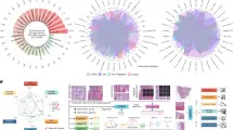
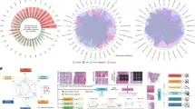
Addressing this critical gap, a multidisciplinary team of researchers has introduced Multi-OSCC, a pioneering, large-scale histopathology dataset designed specifically for multi-target analysis in OSCC. Featuring 1,325 high-resolution images from 1,325 patients captured across multiple magnifications ($times 200$, $times 400$, and $times 1000$), Multi-OSCC uniquely bridges the gap between pathology and clinical outcomes by supporting six distinct diagnostic and prognostic tasks. This breakthrough initiative promises to accelerate the evolution of holistic, AI-assisted clinical decision-making.
Detailed Chronology and Methodological Framework
The journey toward developing the Multi-OSCC benchmark reflects a rigorous, methodical approach to clinical data curation, image acquisition, and computational validation.
Retrospective Cohort Assembly and Governance
Conducted in strict compliance with the Declaration of Helsinki, the retrospective study received institutional review and formal approval from the Ethics Committee of Sun Yat-sen Memorial Hospital, Sun Yat-sen University (Approval Number: SYSKY-2024-440-01). Because the study relied on retrospective clinical records and de-identified archives without compromising personal privacy or commercial interests, an exemption for informed consent was granted.
Patient recruitment targeted individuals who underwent primary surgical resection for pathologically confirmed OSCC at the institution between 2015 and 2022. Inclusion criteria mandated that each patient possessed at least two years of post-operative follow-up data—a crucial timeframe given that the vast majority of OSCC recurrences manifest within the initial 24-month post-surgical window. Notably, the actual follow-up duration for non-recurrence patients significantly exceeded this baseline, averaging 1,404 days to effectively capture potential late-onset recurrence events.
Multi-Magnification Data Acquisition
For every enrolled patient, pathologists prepared two distinct formalin-fixed, paraffin-embedded (FFPE) tissue blocks: one sampled directly from the central core of the tumor and the other from the invasive peripheral margin. These blocks were sectioned and stained with hematoxylin and eosin (H&E). Expert pathologists identified high-yield diagnostic regions of interest exhibiting hallmark structural features, such as cellular differentiation patterns, epithelial arrangements, and cancer nests.
Utilizing advanced microscopy, high-resolution digital images ($2592 times 1944$ pixels) were captured from both core and edge slides across three distinct magnification levels ($times 200$, $times 400$, and $times 1000$). This comprehensive imaging protocol yielded six distinct histopathological views per patient, capturing essential cytoarchitectural components including individual cancer cells, keratin pearls, and nuclear atypia. Clinicopathological annotations and time-to-event timelines were concurrently extracted from the hospital’s electronic medical records (EMR).
Computational Pipeline and Model Architecture
To process this high-dimensional multi-slice data, the research team engineered a modular computational pipeline featuring three core components:
- Pre-trained Vision Backbones: The team evaluated various Convolutional Neural Networks (CNNs) and Vision Transformers (ViTs)—including ResNet50, DenseNet121, ViT-Base, Swin-Base, and pathology-specific foundations like PathoBench, Hibou-B, and CONCH. Pathology-specific pre-training (specifically ViT-Small with PathoBench pre-training) consistently outperformed ImageNet-initialized weights, delivering superior feature extraction capabilities.
- Feature Fusion Modules: To integrate the six feature vectors extracted per patient, the team tested multiple strategies, including Concatenation (Concat), Low-rank Multimodal Fusion (LMF), Gated Fusion, and Transformer-based cross-attention. Simple feature concatenation yielded the highest mean Area Under the Curve (AUC) of 75.00% across all tasks while maintaining architectural simplicity.
- Multi-Task Learning Framework: To handle simultaneous predictions, the team implemented a hard parameter sharing paradigm. Optimization strategies included Sum Loss, GradNorm, and Pareto optimization, with GradNorm proving most robust for balancing gradient magnitudes across tasks.
Supporting Context & Quantitative Metrics
The Multi-OSCC dataset is uniquely structured to power six distinct clinical endpoints, divided into a prognostic task and five granular diagnostic tasks:
- Recurrence (REC): A prognostic metric predicting the two-year risk of post-surgical tumor recurrence.
- Lymph Node Metastasis (LNM): A diagnostic classification aiding decisions regarding neck dissection procedures.
- Tumor Differentiation (TD), Tumor Invasion (TI), Cancer Embolus (CE), and Perineural Invasion (PI): A battery of microscopic evaluations providing deep assessments of tumor aggressiveness and local tissue involvement.
Comprehensive Benchmark and Experimental Findings
Extensive validation experiments generated robust performance baselines across the dataset:
- Stain Normalization Impact: Evaluating Reinhard, Vahadane, and Macenko normalization techniques revealed a fascinating task dependency. While stain normalization significantly enhanced performance on the five diagnostic tasks (with the Reinhard method emerging as the most effective), it impaired performance on the REC task, indicating that original color information holds critical, uncaptured prognostic signals. Consequently, a hybrid approach was adopted—applying Reinhard normalization to diagnostic tasks while preserving raw color data for recurrence prediction.
- Multi-Slice Integration: Combining tissue slices from both the tumor core and invasive margin (Core + Edge) yielded a superior mean AUC of 75.00%, outperforming models restricted to a single tissue region. This confirms that integrating spatial heterogeneity enhances predictive power.
- Pathology versus Clinical Data: Evaluating EMR variables using ClinicalBERT yielded a mean AUC of 61.99%, whereas pathology-based models achieved a mean AUC of 73.59%. For the REC task specifically, the pathology model achieved an outstanding AUC of 93.13%, dwarfing the EMR-only model’s AUC of 76.94%. This demonstrates that micro-morphological phenotypes encode profound signals regarding systemic tumor behavior.
Official Statements and Expert Insights
Principal investigators and clinical collaborators emphasized the transformative implications of the Multi-OSCC benchmark for computational pathology and clinical oncology.
Lead supervisors of the research initiative noted:
"For too long, artificial intelligence in oral oncology has been bottlenecked by fragmented, single-task datasets that mirror neither the complexity nor the integrated nature of actual clinical workflows. By uniting multi-magnification imaging with both diagnostic and prognostic targets within a single, large-scale patient cohort, Multi-OSCC provides the foundational bedrock necessary to move AI tools from experimental novelties into trusted clinical assistants."
Pathologists involved in the manual annotation and Grad-CAM++ interpretability validation added:
"When we reviewed the model’s attention heatmaps—such as its focus on keratin pearls for tumor differentiation or infiltrated striated muscle during invasion analysis—we observed a striking alignment with human expert reasoning. The AI is not merely finding statistical shortcuts; it is learning to recognize the true biological hallmarks of cancer aggressiveness."
Future Outlook and Data Availability
While Multi-OSCC represents a monumental step forward, the authors candidly acknowledge certain limitations. The current dataset relies on high-resolution image patches rather than whole slide images (WSIs). Although patch sampling across core and edge regions effectively captures local cellular morphology, it cannot fully encapsulate the global spatial architecture achievable via complete WSIs.
Future research initiatives will focus on bridging the gap between patch-level cytological detail and WSI-level global context, paving the way for spatially aware, multimodal AI architectures that integrate genomics, clinical EMR data, and comprehensive digital pathology.
Data and Code Availability
To foster transparency, collaboration, and reproducible research, the complete Multi-OSCC dataset (comprising 34.6 GB of compressed high-resolution images, metadata JSON files, and clinical CSV spreadsheets containing time-to-event data) is publicly available via Zenodo (DOI: 10.5281/zenodo.16842637).
Furthermore, all source code for model training, benchmarking protocols, stain normalization scripts, and evaluation metrics has been released on GitHub under the repository guanjinquan/OSCC-PathologyImageDataset.
Through the release of Multi-OSCC, the research community is well-equipped to accelerate the development of holistic, reliable, and clinically actionable artificial intelligence systems designed to improve patient survival and redefine the standard of care in oral oncology.