








Executive Overview
What prompts an individual to cross a room, strike up a conversation, or reach out to another living being? While centuries of philosophy, psychology, and modern neuroscience have sought to map the complex web of human socialization, the exact physical triggers that convert ambient social stimuli into a deliberate decision to approach remain elusive. Now, groundbreaking research emerging from the Hebrew University of Jerusalem is rewriting our understanding of social initiation.
According to a pivotal new study conducted at the Edmond and Lily Safra Center for Brain Sciences (ELSC), the neurological framework for a social approach begins unfolding within the brain several seconds before any physical movement takes place. Spearheaded by Dr. Lilah Avitan alongside PhD student Imri Lifshitz and a multidisciplinary team of laboratory researchers, the study reveals that the intention to socialize is preceded by a distinctive, brain-wide pattern of neural activity.
By observing transparent organism models in real time, the research team discovered that this distributed neural signature does more than just herald an impending movement; its very intensity correlates directly with an individual’s underlying social drive. These insights offer unprecedented windows into why some organisms—and by extension, humans—are naturally gregarious, while others tend toward isolation. More importantly, because fundamental brain-wide organizational patterns are conserved across species, this discovery establishes a critical baseline for exploring human social mechanics, as well as the neural disruptions underlying clinical conditions characterized by altered social functioning, such as autism spectrum disorders and severe social anxiety.
Detailed Chronology & Methodology: Tracking Social Decisions in Real Time
To capture the fleeting neurological moments that precede a social action, the research team at the Hebrew University of Jerusalem faced a formidable technological hurdle. Traditional neuroimaging tools often lack the spatial resolution required to track individual neurons across an entire brain simultaneously, or they are too cumbersome to record split-second behavioral shifts in real time.
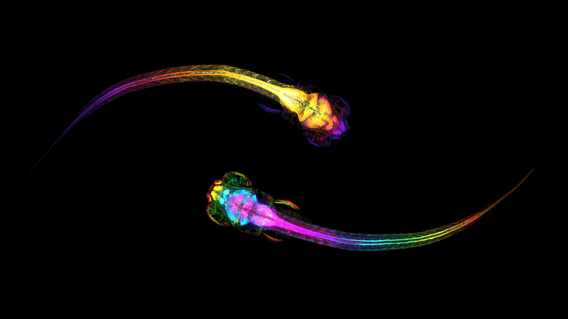
To circumvent these limitations, the laboratory utilized zebrafish (Danio rerio), an established model organism in modern neuroscience. Zebrafish offer a unique combination of physiological advantages: during their early developmental stages, their bodies are nearly transparent, allowing scientists to monitor real-time cellular and sub-cellular brain activity using advanced optical imaging techniques without invasive procedures. Furthermore, their neural architecture shares deep evolutionary homology with more complex vertebrates, including mammals.
Engineering a Dynamic Social Environment
Rather than studying the zebrafish in isolation or exposing them to static visual cues, the researchers engineered an innovative experimental paradigm designed to mimic naturalistic social dynamics.
- The Observation Chamber: The setup placed an "observer" zebrafish in a controlled micro-environment where it could visually track and respond to a conspecific (another zebrafish) swimming freely in an adjacent compartment.
- Brain-Wide Calcium Imaging: Utilizing state-of-the-art light-sheet fluorescence microscopy, the team recorded the neural activity of the observer fish across its entire brain simultaneously. By employing genetically encoded calcium indicators—proteins that fluoresce brightly when a neuron fires—the researchers could visualize the firing patterns of individual cells as they processed real-time social information.
- High-Resolution Behavioral Tracking: Simultaneously, high-speed cameras tracked the precise physical movements of the observer fish, cataloging every twitch, turn, and forward propulsion.
By synchronizing the high-resolution optical brain data with precise behavioral tracking, the research team was able to perform a backward temporal reconstruction. They could trace the neural events leading up to a specific social decision—such as the exact moment a fish decided to swim toward its neighbor—and analyze how the brain orchestrated this action second by second.
A Brain-Wide Signal Appears Before Social Behavior
The temporal analysis yielded remarkable results. When an observer fish prepared to initiate a social approach toward its neighbor, the underlying neurological changes did not happen concurrently with the movement; instead, they preceded the physical action by several seconds.
This revelation challenges reductionist views of brain function. For decades, traditional models of behavioral neuroscience often searched for isolated "centers" in the brain responsible for specific social drives. However, the Hebrew University study demonstrates that social approach behavior is not managed by a single isolated module.
The Neural "Pre-Decision State"
Instead, the decision to socialize is mediated by a coordinated, brain-wide symphony of excitation and inhibition:
- Pallial Activation: The researchers observed a pronounced increase in neural activity within the pallium, a higher-order brain structure in fish that is evolutionarily and functionally homologous to the mammalian cortex and telencephalon. The pallium is traditionally known to process complex cognitive functions, contextual learning, and goal-directed behaviors.
- Widespread Suppression: Concurrently, the team noted a systematic decrease in neural activity across several other distinct subregions of the brain.
- Predictive Architecture: Together, these synchronized increases and decreases formed a distinct spatial-temporal profile—a neurological "pre-decision state." This brain-wide pattern acted as an internal harbinger, signaling reliably that a social interaction was imminent.
Because this signature manifested well before the fish executed the physical stroke of its tail, the researchers could use the strength and configuration of this neural pattern to accurately predict both the timing and the nature of the upcoming behavior. The brain, it appears, commits to a social interaction internally long before the body ever sets the plan into motion.
Supporting Context, Metrics, and Individual Variations
One of the most intriguing dimensions of the study is the discovery that this pre-decision neural signature is not uniform across all subjects. When the researchers compared the neurological data across multiple individual fish, they uncovered significant variance in the strength of the brain-wide pattern.
Quantifying Social Drive
By correlating the intensity of the neural "pre-decision state" with the subjects’ subsequent behavioral choices, the team identified a direct proportionality:
- High-Drive Individuals: Fish that exhibited a robust, highly synchronized brain-wide pattern consistently demonstrated a higher frequency of social approach behaviors. They spent more time near the adjacent partition, displayed heightened attentiveness to the neighboring fish, and initiated contact more rapidly.
- Low-Drive Individuals: Conversely, subjects that displayed weaker or more fragmented pre-decision signatures exhibited significantly lower baseline social motivation, frequently choosing to swim away from the partition or remaining indifferent to the presence of the neighboring fish.
This quantitative link suggests that the neural signature acts as a direct proxy for an individual’s internal social drive. It provides a biological explanation for why two individuals placed in the exact same environment with identical stimuli can make radically different behavioral choices: their brains are generating internal motivational states of varying intensities.
Furthermore, the data reaffirmed the critical importance of the pallium. The intensity of the signals originating within this higher-order region was the single best predictor of an individual’s overall social engagement, cementing its role as the primary neurological engine driving social motivation.
Official Statements and Expert Insights
The implications of these findings have resonated deeply within the global neuroscience community, bridging cellular biology with behavioral psychology.
Highlighting the dual predictive power of the discovery, lead researcher Dr. Lilah Avitan noted:
"This study identifies a brain-wide neural signature of social approach that emerges before movement begins. This signature predicts not only whether an upcoming action will be social, but also how strongly socially driven the individual is."
Dr. Avitan’s remarks underscore a fundamental shift in how neuroscientists view internal states. Rather than treating motivation as a vague psychological construct, the research demonstrates that motivational drive has a concrete, measurable, and predictive neurological footprint that can be tracked across the brain in real time.
Co-authors and institutional representatives at the Edmond and Lily Safra Center for Brain Sciences (ELSC) emphasize that the success of the study hinges on its integrative methodology. By combining whole-brain cellular imaging with naturalistic behavioral paradigms, the laboratory has bridged a longstanding gap between microscopic neural circuit analysis and macroscopic behavioral outcomes.
Future Outlook: Implications for Human Health and Neurology
While the foundational experiments of this research were conducted using zebrafish models, the translational implications for human health and clinical neurology are profound.
Bridging Animal Models and Human Behavior
The fundamental neuroanatomical structures involved in decision-making, motivation, and social interaction—such as the telencephalon, pallium, and associated limbic pathways—share deep evolutionary roots across vertebrates. Consequently, understanding how a simpler vertebrate brain compiles sensory data, generates a pre-decision motivational state, and executes social approach behavior provides an invaluable template for studying more complex mammalian systems.
Addressing Disorders of Social Function
Many psychiatric and neurodevelopmental conditions are defined by an atypical capacity for social engagement. Conditions such as:
- Autism Spectrum Disorder (ASD): Often characterized by differences in social communication, processing of social cues, and intrinsic social motivation.
- Social Anxiety Disorder: Involves an hyperactive or dysregulated anticipatory threat response that inhibits social approach behaviors.
- Schizophrenia: Frequently features severe deficits in social cognition, emotional resonance, and motivation to interact with others.
By identifying the specific neural signatures and brain-wide coordination required to transition from isolation to social engagement, neuroscientists can begin to pinpoint where these pathways malfunction in clinical contexts.
The Road Ahead
As the research team at the Hebrew University of Jerusalem looks toward the future, the next phases of investigation will likely explore how these pre-decision neural states are modulated by environmental stressors, learning experiences, and pharmacological interventions. Researchers aim to determine whether these neural signatures are plastic—meaning they can be trained, altered, or rehabilitated—opening up new theoretical avenues for therapeutic interventions in humans.
Ultimately, this study transforms our perspective on the anatomy of connection. It reminds us that every handshake, every conversation, and every friendly approach is the culmination of a sophisticated, beautifully coordinated neurological overture—one that begins playing in the quiet architecture of the brain long before we ever take a step toward one another.