








Executive Overview
In a scientific breakthrough that fundamentally challenges a foundational dogma of modern immunology, an international team of researchers has uncovered an entirely unrecognized mechanism by which the human immune system can hunt down and destroy cancer cells. Published in the prestigious journal Nature Immunology, the study rewrites our understanding of how white blood cells interact with malignant growths. For decades, the biomedical consensus maintained a strict functional division within the adaptive immune system: Major Histocompatibility Complex (MHC) class I molecules communicated exclusively with CD8+ "killer" T cells, while MHC class II molecules activated CD4+ "helper" T cells. This black-and-white framework has driven decades of cancer research, shaped the design of immunotherapies, and informed the management of post-transplant complications.
However, this new collaborative study—led by Dr. Pavan Reddy, director of the Dan L Duncan Comprehensive Cancer Center at Baylor College of Medicine (BCM), alongside Dr. Arul Chinnaiyan and Dr. Marcin Cieslik of the University of Michigan Rogel Cancer Center—demonstrates that reality is far more flexible and complex. The researchers have proved that the MHC class I pathway plays a vital, previously unknown role in immune responses directly driven by CD4+ T cells.
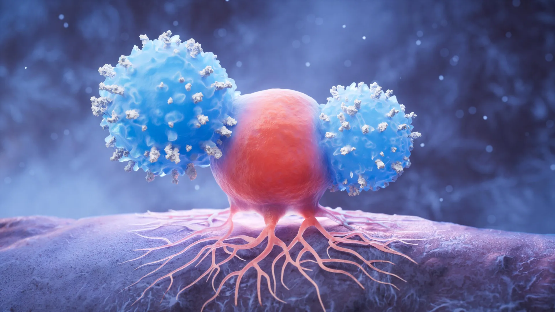
Even more provocatively, the discovery exposes a critical Achilles’ heel in how tumors evade our natural defenses. Many cancers survive by downregulating or entirely shedding their MHC class I surface markers, effectively rendering themselves invisible to killer CD8+ T cells. Yet, this evasive maneuver carries a fatal trade-off: stripping away MHC class I paradoxically leaves these malignant cells wide open to lethal counterattacks from CD4+ T cells. Rather than escaping destruction, the cancer cells trigger a specialized form of iron-dependent cell death known as ferroptosis.
Beyond oncology, these findings hold profound implications for bone marrow transplantation, specifically in understanding and mitigating graft-versus-host disease (GVHD). By illuminating a completely new axis of cellular immunity, this research lays the groundwork for a transformative generation of immunotherapies designed to outsmart drug-resistant tumors and rewrite therapeutic protocols for transplant medicine.
Detailed Chronology: The Path to Paradigm-Shifting Discovery
Unraveling a Decades-Old Biological Dogma
To appreciate the magnitude of the recent findings, one must understand the bedrock principles upon which immunology has rested for over half a century. Since the discovery of MHC molecules in the mid-20th century, textbooks have taught a rigid division of labor. MHC class I proteins are expressed on the surface of virtually all nucleated cells in the human body. Their primary evolutionary purpose is to display internal peptide fragments—including viral proteins or mutated tumor antigens—to passing CD8+ cytotoxic T cells. Upon recognizing these anomalous peptides, the CD8+ T cells execute the target cell via targeted apoptosis. Conversely, MHC class II molecules are restricted primarily to specialized immune cells, presenting extracellular antigens to CD4+ helper T cells, which orchestrate the broader immune response.
This textbook binary—MHC I for CD8+, MHC II for CD4+—became the guiding principle for modern oncology, guiding the development of checkpoint inhibitors, therapeutic cancer vaccines, and cellular therapies. Yet, lingering clinical puzzles suggested that T cell dynamics were far more nuanced than current models could explain.
Recognizing these gaps, a multi-institutional team spearheaded by Dr. Pavan Reddy at BCM and Drs. Arul Chinnaiyan and Marcin Cieslik at the University of Michigan embarked on a rigorous, multi-year investigation. The core research team—including graduate students and key contributors Emma Lauder and Meng-Chih Wu from Baylor, alongside Mahnoor Gondal from the University of Michigan—set out to dissect the molecular dialogue between T cells and target tissues using high-resolution genomic tools.
Pinpointing the Cross-Talk Between MHC I and CD4+ T Cells
The breakthrough began when the researchers started analyzing complex transcriptomic datasets to observe how cells respond when standard immune communication pathways are disrupted. Utilizing advanced functional studies combined with sophisticated murine (mouse) models and human tissue samples, the team systematically mapped out cellular behaviors that traditional immunology models failed to predict.
Through meticulous experimentation, the investigators noticed something extraordinary: when MHC class I expression was experimentally reduced or completely silenced, the targeted cells did not become universally protected from immune clearance. Instead, they experienced a dramatic shift in vulnerability. The absence of MHC I molecules triggered a potent, secondary immune response mediated entirely by CD4+ helper T cells.
This counterintuitive observation completely upended the prevailing paradigm. The researchers realized that the MHC class I pathway—long assumed to be the exclusive domain of CD8+ killer T cells—actively suppresses or modulates CD4+ T cell-mediated cytotoxicity under specific conditions. When that suppression is lifted, CD4+ T cells step into the vacuum, executing a devastatingly effective strike against the target cells.
The Mechanism of Destruction: Unlocking Ferroptosis
Identifying the involvement of CD4+ T cells was only the first step; the team needed to understand how these helper cells were killing target cells that lacked MHC I. Through rigorous biochemical and transcriptomic analyses, the investigators tracked the cellular cascade that follows CD4+ T cell engagement.
The weapon of choice was not traditional apoptosis or necrosis, but ferroptosis—an iron-dependent form of regulated cell death driven by the lethal accumulation of lipid peroxides and oxidative stress. When CD4+ T cells encountered cells with depleted MHC I levels, they initiated a signaling cascade that overwhelmed the target cell’s antioxidant defenses, particularly glutathione peroxidase 4 (GPX4) pathways, leading to unchecked iron-mediated membrane destruction.
For cancer research, this discovery carries monumental irony. Tumors frequently evolve mechanisms to downregulate MHC class I molecules to evade the relentless hunting of CD8+ cytotoxic T cells—a classic immune-evasion tactic known as "HLA loss" or "MHC class I downregulation." However, the Baylor and University of Michigan team demonstrated that this adaptive survival strategy is, in reality, a trap. By hiding from CD8+ cells, the tumor inadvertently rolls out the welcome mat for CD4+ T cell-driven ferroptosis. The cancer cells simply trade one executioner for another, more unexpected one.
Supporting Context & Metrics: Broad Implications in Oncology and Transplantation
Bridging Solid Tumors and Checkpoint Inhibitor Outcomes
To ensure that these laboratory discoveries were not merely academic anomalies confined to mouse models, Dr. Chinnaiyan’s computational pathology team at the University of Michigan undertook a massive translational data analysis. They examined extensive transcriptomic and clinical outcome datasets from human patients who had undergone checkpoint inhibitor immunotherapy for various solid tumors.
The computational analysis yielded striking correlations. Patients whose tumors exhibited altered or lowered MHC class I expression profiles showed distinct clinical response patterns when treated with immunotherapies that mobilize T cell responses. These findings confirmed that the newly discovered MHC I-CD4+ ferroptosis axis operates in human clinical settings, directly influencing how patients respond to modern cancer treatments.
These metrics suggest that tumors previously classified as "immunologically cold" or resistant to standard immunotherapies because of MHC I downregulation may actually be primed for alternative forms of immune destruction. By designing therapies that intentionally manipulate or exploit this pathway, clinicians may soon be able to reactivate stalled immune responses in stubborn cancers.
Beyond Oncology: Graft-Versus-Host Disease (GVHD)
The scope of the discovery extends far beyond oncology into the delicate and high-stakes realm of hematopoietic stem cell transplantation (HSCT), commonly known as bone marrow transplantation. During bone marrow transplants, a major life-threatening complication is graft-versus-host disease (GVHD), wherein the donor’s immune cells (the graft) attack the recipient’s healthy tissues (the host).
When the research team expanded their investigation into models of transplantation, they observed that the exact same ferroptosis-driven, CD4+ T cell-mediated mechanism was at play in allogeneic tissue damage. Just as low MHC class I expression makes cancer cells vulnerable to CD4+ attacks, it also sensitizes healthy host tissues to immune-mediated destruction during GVHD.
This dual relevance makes the discovery exceptionally powerful. By understanding how MHC class I regulates tissue sensitivity to CD4+ T cell damage, scientists can now pursue dual-purposed therapeutic strategies: finding ways to harness this pathway to obliterate drug-resistant tumors while simultaneously developing protective interventions to shield vital patient tissues from catastrophic post-transplant complications.
Official Statements and Collaborative Vision
The breadth of this discovery reflects the power of deep, multidisciplinary collaboration across premier academic medical institutions. Dr. Pavan Reddy emphasized the far-reaching potential of the findings during statements discussing the publication in Nature Immunology.
"Our work, if further validated, will have implications for T cell-mediated immune responses beyond cancer and transplant immunology," stated Dr. Reddy, director of the Dan L Duncan Comprehensive Cancer Center at Baylor College of Medicine. "This may allow for the development of novel strategies that target MHC class I and CD4+ T cells to leverage the beneficial side of immunity or mitigate unwanted immune responses."
The research project represents a triumph of cross-institutional teamwork, drawing on the clinical oncology expertise of Baylor College of Medicine and the cutting-edge pathological and computational capabilities of the University of Michigan Rogel Center. The foundational investigative work was propelled forward by a dedicated cohort of graduate students and postdoctoral researchers, including Emma Lauder, Meng-Chih Wu, Mahnoor Gondal, Akira Yamamoto, Laure Maneix, Dongchang Zhao, and Yaping Sun. Their combined expertise bridged mouse genetics, human tissue pathology, single-cell transcriptomics, and advanced biostatistical modeling.
This ambitious research agenda has been sustained by robust financial backing from federal agencies and philanthropic research organizations. The study received vital funding through grants from the National Institutes of Health (NIH)—including project numbers P01CA039542, P01HL149633, R01HL152605, R01CA217156, R01AI165563, CA125123, OD036336, and OD038251—as well as vital support from the Cancer Prevention and Research Institute of Texas (CPRIT) via grants RR220033 and RP240432. Additional institutional support was provided through affiliations with the Howard Hughes Medical Institute.
Future Outlook: The Next Frontier in Immunotherapy
As the medical community digests the implications of this Nature Immunology publication, translational researchers are already looking toward the horizon. The transition from basic biological discovery to clinical application is complex, but the potential clinical dividends are immense.
Designing Next-Generation Immunotherapies
Current immunotherapies—such as CAR-T cell therapies and immune checkpoint blockades (anti-PD-1/PD-L1 and anti-CTLA-4)—are primarily engineered to enhance the activity of CD8+ killer T cells or unleash existing immune responses. However, a significant percentage of patients experience therapeutic failure because their tumors acquire resistance through the downregulation of MHC class I molecules.
Armed with the knowledge that MHC I-deficient cancer cells are susceptible to CD4+ T cell-mediated ferroptosis, future drug development can focus on:
- Combination Therapies: Formulating treatments that simultaneously block immune checkpoints while therapeutically inducing lipid peroxidation or iron accumulation in tumor cells, thereby weaponizing CD4+ T cells against MHC I-low cancers.
- Targeted CD4+ Mobilization: Developing vaccines or adoptive T cell therapies specifically designed to expand and direct CD4+ helper cells toward pathways that trigger ferroptosis in treatment-resistant malignancies.
Refining Bone Marrow Transplantation Protocols
In transplant immunology, the discovery provides a novel therapeutic target for controlling graft-versus-host disease. By identifying how MHC class I expression modulates host tissue susceptibility to CD4+ T cell attacks, researchers can investigate pharmacologic agents that inhibit ferroptosis in healthy recipient tissues during the acute post-transplant window. This could decouple the beneficial "graft-versus-tumor" (GVT) effect from the destructive "graft-versus-host" pathology, making bone marrow transplants significantly safer for leukemia and lymphoma patients.
Validation and Clinical Trials
Before these concepts reach the bedside, the scientific team emphasizes that rigorous further validation is required. Preclinical testing in advanced humanized mouse models and retrospective analyses of clinical trial cohorts are already underway to optimize therapeutic dosages and identify reliable biomarkers. These biomarkers will help clinicians predict which patients are most likely to respond to therapies targeting the MHC I-CD4-ferroptosis axis.
Ultimately, the Baylor and University of Michigan study stands as a powerful reminder of how much remains undiscovered within human biology. By challenging a decades-old consensus, these researchers have not only solved a persistent immunological mystery but have also opened a promising new chapter in the ongoing war against cancer and transplant complications.