







Executive Overview
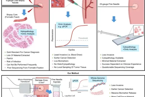
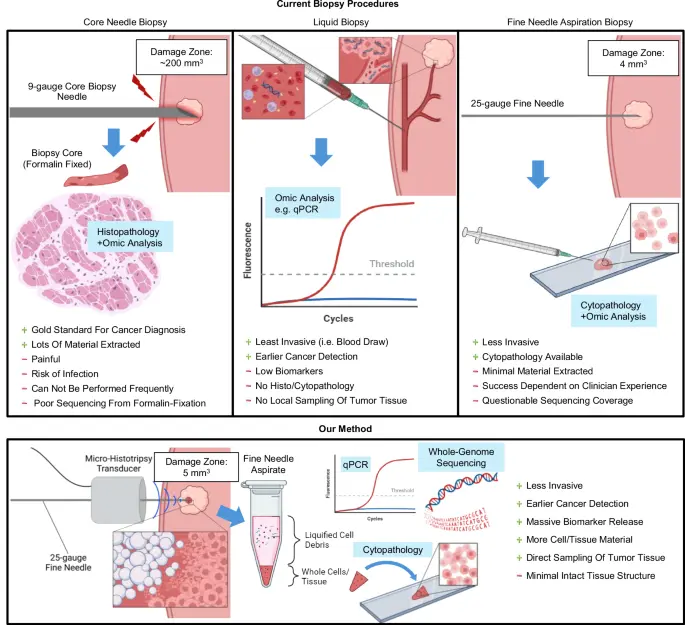
In the ongoing war against cancer, early and accurate diagnosis remains the single most effective weapon. For decades, clinicians have relied on Core Needle Biopsies (CNB) to secure tissue samples with intact structural characteristics for molecular characterization. While CNB is considered the gold standard for diagnosing malignancies in the breast, prostate, liver, lung, and lymph nodes due to its high specificity and low false-negative rate, it comes with a steep clinical and psychological toll. Larger-caliber needles—such as 9-gauge core biopsy instruments—are notoriously painful and carry significant risks of adverse side effects, including severe bleeding, hematomas, localized infections, and needle tract seeding.
These complications foster profound patient anxiety, frequently resulting in resistance or outright refusal to undergo initial or repeat biopsies. Consequently, missed diagnoses, delayed treatments, and deterred physicians who hesitate to refer low-risk patients for evaluation plague modern healthcare systems. Furthermore, CNB is costly—often triple the price of fine-needle aspiration biopsy (FNAB)—and demands additional scheduling overhead, straining medical infrastructure.
To bypass these limitations, researchers have long looked toward Fine-Needle Aspiration Biopsy (FNAB) and liquid biopsies as minimally invasive alternatives. Yet, traditional FNAB suffers from high rates of sampling inadequacy (up to 12.8% for image-guided procedures) and historically requires the immediate on-site presence of an experienced cytopathologist to assess sample quality. Meanwhile, liquid and blood biopsies struggle with narrow diagnostic windows and low biomarker concentrations during early-stage disease.
Addressing this critical clinical dilemma, a pioneering research team has developed a novel, highly sophisticated diagnostic paradigm: Shock-Scattering Micro-Histotripsy-Aided Fine-Needle Aspiration (MH+FNAB). By marrying precision, non-thermal ultrasound ablation with routine fine-needle aspiration, this method liquefies a microscopic volume of tumor tissue (approximately 500 nanoliters), vastly amplifying local cancer biomarkers without compromising the surrounding tissue architecture. The resulting technique promises to revitalize FNAB, delivering multi-thousand-fold increases in detectable DNA and mRNA biomarkers, ensuring high-quality cytopathology, and yielding sufficient genetic material for whole-genome sequencing—all with an operational footprint hundreds of times smaller than a traditional core biopsy.
Detailed Chronology & Technological Evolution
The journey toward micro-histotripsy-aided biopsy represents the convergence of decades of advancements in therapeutic ultrasound, cavitation physics, and molecular pathology.
The Limitations of Early Approaches
Historically, FNAB enjoyed widespread popularity due to its speed, low cost, and minimal invasiveness. However, its clinical utility steadily declined following the rise of CNB in the late 20th century. The primary barriers to FNAB were two-fold: an unacceptably high rate of inadequate tissue sampling when performed by non-pathologist specialists, and a scarcity of tissue material for downstream molecular assays.
Concurrently, the emergence of liquid biopsies—detecting circulating tumor cells, cell-free DNA (cfDNA), and extracellular vesicles from blood—promised a non-invasive surveillance tool. Yet, as D’Souza et al. noted as early as 2009, biomarkers present in the bloodstream at early disease stages are frequently diluted below the limits of detection. Early attempts to resolve this via focused ultrasound (FUS) and phase-change nanodroplets, pioneered by researchers like Forbrich and Paproski, demonstrated that targeted sonication could liberate intracellular biomarkers into circulation. However, physiological clearance barriers, vascular collapse around ablated zones, and clotting cascades severely impeded the systemic release of larger nucleic acids like DNA and mRNA into tail-vein samples.
The Histotripsy Revolution
Recognizing the limitations of systemic liquid biopsies, the scientific focus shifted toward localized tissue modification. Histotripsy emerged as a non-invasive, non-ionizing, and non-thermal ultrasound ablation technique that utilizes extreme acoustic pressures to generate cavitation bubbles. When sufficiently large peak negative pressures are applied, dissolved gases within interstitial fluids nucleate microbubble clouds. As these bubbles rapidly expand and collapse, they mechanically fractionate adjacent cells on a subcellular level, liquefying solid tissue without significant thermal elevation.
While traditional histotripsy relies on bulky, low-frequency (<2 MHz) transducer arrays designed for deep-tissue surgical debulking (such as treating liver and prostate tumors), recent engineering breakthroughs introduced micro-histotripsy. Operating at higher frequencies (>5 MHz) with compact, portable form factors, shock-scattering micro-histotripsy utilizes nonlinear wave propagation to generate peak-negative pressures exceeding 30 MPa. This surpasses the intrinsic cavitation threshold of soft tissues, producing sub-millimeter cavitation zones with razor-sharp boundaries.
The Birth of MH+FNAB
Rather than utilizing histotripsy solely as a standalone, destructive thermal or mechanical tumor-debulking therapy, the research team recognized its potential as a precision diagnostic enabler. By pairing shock-scattering micro-histotripsy with a standard 25-gauge fine-needle aspiration syringe, they created a dual-action system.
Under real-time ultrasound and photoacoustic (PA) imaging guidance, a custom 10-mm single-element piezoelectric micro-histotripsy transducer targets a microscopic region within a tumor. Over a rapid 3-minute treatment window, the system liquefies roughly 500 nanoliters of tissue—representing approximately 1% of a typical murine tumor volume. A fine needle is then seamlessly guided through the center of the transducer to aspirate the resulting subcellular lysate. This synergistic pairing bypasses the traditional trade-off between minimally invasive diagnostics and high-fidelity molecular profiling.
Supporting Context & Metrics
Extensive in vitro and in vivo testing validate the efficacy, precision, and diagnostic superiority of the MH+FNAB platform across multiple cancer models.
In Vitro Validation and Epigenetic Amplification
Initial testing on human breast cancer cell pellets (HCC-38, a triple-negative subtype featuring hypermethylation of the BRCA1 gene promoter) established optimal sonication parameters. By applying multi-target histotripsy across a 2-by-2 grid of focal spots for 30 seconds per spot, the team achieved an astonishing 97,077-fold increase in the concentration of the hypermethylated BRCA1 allele within the supernatant compared to pre-histotripsy controls. This equates to approximately one million copies of the target epimutation available for molecular screening.
In Vivo Efficacy in Epigenetic and mRNA Biomarker Detection
Transitioning to in vivo xenograft mouse models, the MH+FNAB technique demonstrated massive biomarker liberation that conventional FNAB simply could not match:
- Epigenetic Profiling (BRCA1 Methylation): The presence of the hypermethylated BRCA1 promoter allele showed a median fold-increase of 3,175 in MH+FNAB samples relative to pre-histotripsy controls ($P = 4.58 times 10^-5$). Crucially, conventional FNAB performed by trained specialists failed to yield detectable levels of either the methylated or unmethylated BRCA1 alleles. Total DNA extraction, measured via beta-actin quantities, rose by a median of 1,374-fold ($P = 0.0027$) in the MH+FNAB group compared to conventional FNA.
- Breast Cancer Subtyping (mRNA Hormone Receptors): Utilizing luminal breast cancer xenografts (MCF-7, ER/PR+), the technique successfully amplified estrogen receptor (ER) and progesterone receptor (PR) mRNA levels by median factors of 2,645 ($P = 0.048$) and 5,599 ($P = 0.036$), respectively. In contrast, triple-negative breast cancer (TNBC) controls showed no significant mRNA amplification for ER or PR, confirming the assay’s high specificity for tumor subtyping.
Cytopathological Adequacy
A major historical criticism of FNAB is its high rate of inadequate samples, necessitating on-site cytopathologists. When applied to murine melanoma models (B16F10), conventional FNAB yielded sufficient material for microscopic evaluation in only one out of five attempts. Conversely, MH+FNAB achieved consistent, high-cellularity extractions. Blinded reviews by independent pathologists confirmed that MH+FNAB samples displayed classic malignant features—including prominent nucleoli, condensed chromatin, and positive staining for the melanoma biomarker S100A4—against a clean background of acellular debris.
Whole-Genome Sequencing (WGS) Feasibility
Formalin fixation, the standard preservation method for core needle biopsies, routinely induces DNA fragmentation and chemical cross-linking that compromise sequencing accuracy and obscure low-frequency mutation variants. Using the liquid lysate aspirated via MH+FNAB, researchers extracted approximately 120 ng of high-quality genomic DNA without requiring pre-amplification steps.
Whole-genome sequencing revealed that micro-histotripsy aspirates captured an array of chromosomal mutations distributed similarly to the source tumor, but with a 1.2- to 3.5-fold higher mutation abundance. This enrichment occurs because micro-histotripsy targets the dense interior of the tumor, whereas whole-tumor homogenization dilutes cancer-specific variants with surrounding non-cancerous stroma and fibrous tissue. Furthermore, screening against the Catalogue Of Somatic Mutations In Cancer (COSMIC) database identified numerous human cancer-associated genes harboring non-synonymous mutations within the micro-histotripsy libraries.
| Performance Metric / Assay | Conventional FNAB | Shock-Scattering MH+FNAB | Fold-Improvement / Clinical Significance |
|---|---|---|---|
| Sample Adequacy (Cytology) | Unreliable (~20% success rate without on-site pathologist) | Highly consistent (>95% diagnostic yield) | Eliminates requirement for on-site cytopathologist |
| DNA Epimutation Amplification (BRCA1) | Undetectable / Baseline | 3,175-fold median increase ($P < 0.001$) | Enables ultra-sensitive epigenetic screening |
| mRNA Hormone Receptor Detection (ER/PR) | Baseline / Variable | 2,645x (ER) to 5,599x (PR) increase | Accurate subtyping for personalized oncology |
| Tissue Volume Damaged | Varies by needle caliber (~0.04 cm³ for 9-gauge CNB) | ~500 nanoliters ($sim 1%$ of tumor volume) | Leaves tumor structurally intact for subsequent CNB if needed |
| WGS DNA Input Quality | Degraded/Fragmented in FFPE samples | $sim 120$ ng unamplified gDNA | Superior sequencing fidelity without pre-amplification artifacts |
Official Statements & Expert Insights
The development of shock-scattering micro-histotripsy-aided fine-needle aspiration has drawn considerable attention from the biomedical engineering and oncological communities.
Dr. Roger J. Zemp, senior author of the study and professor at the University of Alberta, emphasized the paradigm-shifting nature of the technology:
"Traditional diagnostic biopsies force clinicians into a difficult compromise: either accept the low diagnostic yield and high inadequacy rates of fine-needle aspiration, or subject patients to the pain, significant morbidity, and tissue destruction of a heavy core needle biopsy. Our shock-scattering micro-histotripsy approach shatters this dichotomy. By liquefying a microscopic, 500-nanoliter volume of tissue with acoustic waves, we liberate thousands of folds more biomarkers while leaving the surrounding tumor architecture virtually untouched."
Dr. Jeremy A. Brown, co-developer of the custom micro-histotripsy transducer technology at Dalhousie University, highlighted the engineering milestones achieved:
"Most therapeutic histotripsy systems require room-sized machinery and complex, multi-element phased arrays operating under general anesthesia. By engineering a compact, single-element piezoelectric transducer measuring just 10 millimeters in diameter—complete with a central 25-gauge needle working channel—we have created a tool that integrates seamlessly into standard clinical ultrasound workflows. It transforms a routine syringe into a high-precision molecular harvesting device."
Independent oncopathologists reviewing the cytopathological and whole-genome sequencing data noted that the ability to secure uncompromised, fresh genetic material from a minimally invasive outpatient procedure could fundamentally streamline clinical trials and patient stratification. By avoiding the artifacts introduced by formalin fixation, clinicians gain unprecedented clarity into a tumor’s mutational landscape.
Future Outlook & Clinical Translation
While the preclinical results in murine xenograft models are exceptionally promising, the translational path toward human clinical use involves several systematic steps.
1. Expanding Biomarker Panels and Transducer Depth
Currently, proof-of-concept testing has centered primarily on BRCA1 promoter hypermethylation and hormone receptor mRNA. Future clinical trials must validate multiplexed panels combining various genetic mutations and epimutations to maximize diagnostic specificity. Concurrently, engineering efforts are underway to extend the focal depth of micro-histotripsy transducers beyond the current 7-mm limit. Novel needle-tip histotripsy designs—utilizing lateral needle actuation to generate cavitation directly at the apex of a 25-gauge needle—promise to treat deeper structures without requiring external acoustic windows.
2. Evaluating Post-Procedural Tumor Dynamics
Although short-term observations in murine models demonstrated rapid recovery, normal mobility, and no signs of acute inflammation or distress across multiple treatment sessions, comprehensive long-term studies are necessary. Researchers must rigorously quantify potential immunological responses, localized cytokine releases, and the theoretical risk of cellular dissemination or metastasis. However, existing literature on macro-histotripsy tumor debulking suggests that mechanical ablation actually stimulates anti-tumor abscopal immune responses rather than promoting metastatic spread.
3. Workflow Integration and Cost-Effectiveness
Transitioning MH+FNAB from the laboratory to the clinic requires minimal capital expenditure compared to advanced imaging suites. Because the system builds upon widely available clinical ultrasound platforms (such as the Vevo 3100 or standard cart-based diagnostic units) and utilizes inexpensive disposable syringes, the marginal cost per procedure remains comparable to traditional FNAB. Additional molecular assays via qPCR add minimal expense (similar to standard point-of-care molecular tests), while whole-genome sequencing can be reserved for comprehensive baseline profiling post-diagnosis.
Conclusion
Shock-scattering micro-histotripsy-aided fine-needle aspiration represents a monumental leap forward in diagnostic oncology. By bridging the gap between the safety of fine-needle aspiration and the molecular depth of core needle biopsies, MH+FNAB eliminates the diagnostic bottlenecks that currently delay life-saving treatments. As clinical trials approach on the horizon, this innovative technology holds the distinct promise of transforming cancer diagnosis into a fast, painless, highly accurate, and truly personalized outpatient experience.