







Executive Overview
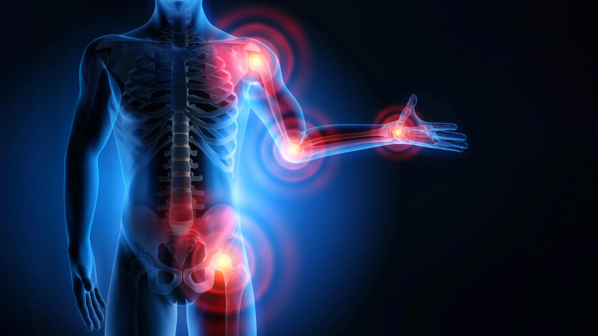
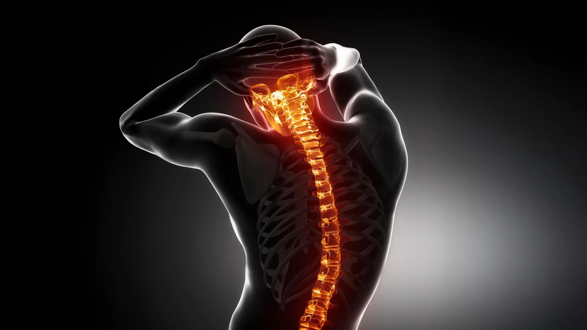
Back pain is a nearly universal human experience, cutting across demographics, lifestyles, and professions. For millions, it is not merely a transient inconvenience but a chronic, debilitating condition that dictates the boundaries of their daily lives. At the epicenter of this global health crisis is intervertebral disc degeneration (IVDD)—the progressive breakdown of the soft, gel-like cushions that act as the spine’s natural shock absorbers. Until recently, medical science has been largely powerless to halt or reverse this structural collapse. For decades, clinicians have had to watch patients deteriorate, relying on symptom management until the structural failure becomes severe enough to warrant invasive spinal surgery.
Now, a groundbreaking collaborative study spearheaded by researchers at the Universities of Edinburgh and Bristol has fundamentally shifted our understanding of how and why our spines break down. Published in the journal Communications Biology and funded by Arthritis UK and the Biotechnology and Biological Sciences Research Council (BBSRC), the research demonstrates that changes in gene activity can directly damage spinal discs by inducing abnormal mineralization. By utilizing an innovative zebrafish model, the scientific team successfully mapped out the biological cascade that causes flexible spinal tissue to turn dangerously hard, resembling misplaced bone growth.
Even more promisingly, the study moves beyond mere observation. By identifying specific biological pathways implicated in the disease—including fat metabolism, phosphate regulation, and growth signaling—the researchers discovered that existing pharmaceutical interventions, such as osteoporosis drugs and dietary adjustments, can dramatically reduce spinal damage in the animal models. This breakthrough not only shatters decades of therapeutic stagnation in spinal medicine but also heralds a potential new era where intervertebral disc degeneration may finally be treated in the doctor’s office rather than the operating theater.
Detailed Chronology: Unraveling the Mechanics of Spinal Degradation
To understand the magnitude of the recent discovery, one must trace the meticulous steps taken by the research teams at Edinburgh and Bristol as they bridged the gap between genetic predisposition and physical degeneration.
The Genetic Link and the Zebrafish Model
For years, genetic studies have hinted that intervertebral disc degeneration does not occur entirely by chance. Epidemiological and genetic research has repeatedly flagged a strong correlation between early-onset disc problems and mutations in genes associated with collagen IX—a vital structural protein responsible for binding together the internal fibrous network of spinal discs. However, proving causality and observing the step-by-step destruction of a living spine in real-time has long eluded scientists.
To crack this physiological puzzle, the researchers turned to an unexpected proxy: the zebrafish. While zebrafish might seem worlds apart from humans anatomically, their spinal columns share fundamental genetic and developmental pathways with our own. Crucially, the research team bred a specialized cohort of zebrafish lacking a functioning copy of the gene linked to collagen IX, allowing them to simulate the genetic defects observed in human disc disease patients.
From Structural Scaffold Failure to Mineralization
As the genetically modified zebrafish aged, the researchers observed a striking phenomenon: the fish developed spinal abnormalities that bore an uncanny resemblance to human IVDD. Their vertebrae began to fuse together, and the soft, compliant tissue acting as the cushion between the bone segments underwent a pathological transformation.
However, the most critical revelation of the study lay in the chronology of this destruction. The mineralization—the process whereby calcium and other minerals precipitate and harden the tissue—did not happen overnight, nor was it the primary event.
Instead, the researchers tracked a distinct, sequential breakdown:
- Scaffolding Collapse: First, a specialized supportive scaffold layer within the developing spine began to structurally deteriorate and weaken.
- Secondary Damage: Following the degradation of this foundational matrix, pathological changes rippled through the microenvironment of the disc.
- Mineral Deposition: Only after this initial structural compromise occurred did mineral deposits begin to aggressively accumulate, turning flexible tissue into rigid, bone-like formations that strangled the spine’s flexibility and fused adjacent vertebrae.
Mapping the Biological Pathways
With the timeline of physical degradation established, the team dove deeper into the molecular engine driving the process. By analyzing genome-wide expression patterns, they sought to answer why the scaffold failed and why minerals rushed in to take its place.
The transcriptomic analysis yielded several critical suspects. The researchers discovered profound disruptions in fat processing pathways and the mTOR signaling pathway, which regulates cell growth and metabolism. Furthermore, they identified aberrations involving phosphate control and vitamin A signaling. Crucially, each of these biological processes has independently been tied to pathological mineral accumulation in other tissues throughout the body, providing a unified mechanistic explanation for how genetic flaws in collagen IX can cascade into widespread spinal hardening.
Supporting Context & Metrics: The Scale of the Back Pain Crisis
The urgency of this research cannot be overstated when viewed against the staggering global burden of back pain.
A Public Health Epidemic
- Universal Prevalence: The vast majority of the human population will experience clinically significant back pain at some point in their lives, making it one of the leading causes of global disability.
- The UK Burden: In the United Kingdom alone, approximately 9.5 million people live with chronic back pain. It is a condition that has plagued families for generations, stripping individuals of their mobility, independence, and economic productivity.
- Economic and Healthcare Strain: The financial toll of IVDD and associated back ailments is astronomical. It accounts for millions of lost workdays, extensive physical therapy regimens, and massive expenditures on diagnostic imaging and surgical interventions.
- The Therapeutic Void: Despite its status as a leading cause of disability worldwide, modern medicine has possessed virtually no disease-modifying drugs for IVDD. Treatments have historically been palliative—consisting of anti-inflammatory medications, physical therapy, and steroid injections—or radical, culminating in spinal fusion surgery or artificial disc replacement when all else fails.
The Pharmacological Breakthrough
Armed with their newfound understanding of the biological pathways driving mineralization, the Edinburgh and Bristol teams tested several interventions to see if the degenerative cascade could be halted or reversed. Their findings open up an immediate pipeline of drug repurposing opportunities:
- Bisphosphonates: By administering bisphosphonates—a class of bone-protecting medications already widely and safely used to treat osteoporosis by inhibiting bone resorption—the researchers successfully prevented abnormal mineral accumulation in the zebrafish spines.
- Metabolic Modulation: Intriguingly, the team discovered that spinal fusion could also be significantly mitigated through lifestyle and metabolic interventions. When the fish received reduced caloric intake or were treated with drugs specifically designed to suppress fat metabolism, the pathological hardening of the spine slowed down considerably.
These results highlight that the biological processes governing phosphate regulation and fat metabolism are not just academic curiosities; they are highly actionable targets for the development of the world’s first disease-modifying medicines for back pain.
Official Statements: Perspectives from the Scientific Vanguard
The implications of the study have drawn enthusiastic responses from academic leaders and the institutional bodies that funded the research, highlighting a shared optimism for the future of spinal medicine.
Dr. Erika Kague, study lead from the University of Edinburgh’s Institute of Genetics and Cancer, captured the historical significance of the breakthrough:
"For decades, surgery has been the only real answer for disc disease. By understanding the biology that drives the spine to harden, our zebrafish studies point to several ways of slowing it down, including a drug already used safely in patients. There’s more work to do, but for a condition that’s affected people for generations without a treatment in sight, this is super exciting."
Dr. Caroline Aylott, Head of Research Delivery at Arthritis UK, emphasized the human impact of the findings for millions of sufferers:
"For the 9.5 million people across the UK living with back pain, this research brings fresh hope that potential new therapeutic approaches are on the horizon. We are proud to fund research that is unlocking the science behind the processes leading to spinal disc degeneration. Back pain is one of the UK’s most common conditions that has blighted millions over generations. Dr. Erika Kague and her team at the University of Edinburgh have uncovered important genetic evidence that could pave the way for new treatments, bringing us one step closer to a future where fewer people have to live with the daily pain and challenges that back pain can bring."
Adding an institutional perspective on the value of foundational science, Dr. Jef Grainger, Executive Director of Bioscience Advancing Knowledge at BBSRC, noted:
"This research shows how publicly funded discovery bioscience can generate the knowledge needed to address major health challenges. By revealing new knowledge of how healthy biological processes break down in aging-related spinal disc degeneration, the study opens up promising avenues for future treatment development. It’s a great example of how BBSRC-supported research helps turn scientific discovery into knowledge and innovations that have the potential to improve people’s lives."
Future Outlook: Toward a Non-Surgical Future for Spine Care
As the scientific community digests the findings published in Communications Biology, the roadmap ahead is clear, albeit demanding. Translating discoveries from aquatic vertebrate models to human clinical applications requires rigorous, phased clinical trials, safety evaluations, and dosage optimizations.
However, the path forward is illuminated by a beacon of hope that has been absent from spinal research for generations. By demonstrating that an existing osteoporosis medication—a bisphosphonate—can successfully thwart the mineralization process in living spines, researchers have bypassed the lengthy and uncertain phases of de novo drug discovery for at least one candidate therapy. Clinical trials exploring the repurposing of bone-protecting drugs for early-stage intervertebral disc degeneration are now within the realm of possibility.
Furthermore, the identification of fat metabolism and phosphate regulation as key drivers of matrix destruction opens the door for pharmaceutical companies to design novel, targeted therapies aimed at preserving the structural integrity of the spinal scaffold before mineralization can take root.
For the millions of individuals worldwide whose lives are currently dictated by the dull ache or sharp agony of a degenerating spine, this study marks the beginning of the end of an era defined by surgical inevitability. As science unlocks the genetic and molecular secrets of our internal shock absorbers, the horizon of medicine grows ever closer to a future where back pain can be intercepted, treated, and managed with a pill rather than a scalpel.