






Executive Overview
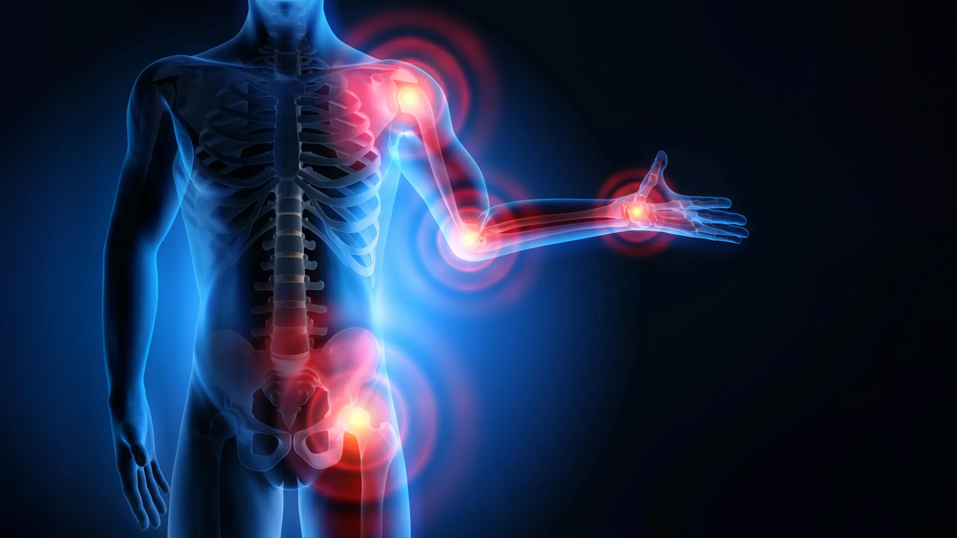
As the global population ages, maintaining musculoskeletal health has emerged as a paramount priority for public health initiatives. While growing older brings wisdom, experience, and the milestones of a life well-lived, it also introduces physical challenges—chief among them being the gradual degradation of joint health. For more than 500 million people worldwide, osteoarthritis and chronic joint discomfort cast a shadow over daily activities, turning routine movements like bending, climbing stairs, or taking a morning walk into sources of friction and pain.
For generations, conventional wisdom erroneously suggested that achy joints meant individuals should rest, avoid physical exertion, and protect their limbs from movement. Modern science, however, has upended this outdated paradigm. Contemporary clinical research demonstrates that targeted, low-impact exercise and intelligent physical conditioning are not only safe for aging joints—they are fundamentally essential. Motion acts as a biological catalyst, lubricating skeletal structures, delivering vital nutrients to avascular tissues, and fortifying the surrounding musculature to act as natural shock absorbers.
This comprehensive report explores the physiological mechanics of joint degradation, examines the transformative clinical data surrounding exercise therapy, evaluates the critical role of proprioception and unstable-surface training, and provides actionable, expert-backed strategies for safely integrating movement into daily life. Whether you are managing early-stage osteoarthritis or seeking to preserve lifelong mobility, understanding the science of joint health offers a powerful pathway to graceful, pain-free aging.
Detailed Chronology: The Biological Mechanics of Joint Aging
To understand why exercise is the ultimate prescription for joint preservation, one must first examine the intricate microarchitecture of the human joint and how it responds to the passage of time.
The Anatomy of Motion: Cartilage and Synovial Fluid
At the intersection of every bone lies a sophisticated biomechanical system. Each joint is cushioned by articular cartilage, a specialized, smooth connective tissue that covers the ends of bones. This remarkable tissue provides a low-friction surface that allows bones to glide effortlessly past one another, while simultaneously distributing mechanical loads evenly across the joint structure.
Surrounding this cartilage is synovial fluid, a viscous, egg-white-like substance housed within the joint capsule. Synovial fluid performs a dual function: it acts as a hydraulic lubricant to minimize friction during movement, and it serves as a vital delivery vehicle, supplying avascular cartilage with critical nutrients.
The Vulnerability of Avascular Tissue
Unlike muscle tissue or bone, which boast robust networks of blood vessels capable of rapid healing and regeneration, articular cartilage lacks its own blood supply. Because cartilage cannot easily repair itself once damaged, cumulative micro-trauma, mechanical stress, and the natural biochemical shifts of aging take a heavy toll.
Over decades, the body experiences a gradual reduction in both the volume of synovial fluid and the density of cartilage-building components. As this protective matrix thaws and thins, bones begin to experience increased friction. This degenerative cascade is the hallmark of osteoarthritis—a systemic condition that progressively erodes joint function, most frequently targeting weight-bearing structures such as the knees, hips, and spine.
The Turning Point: Reframing the Role of Exercise
Historically, individuals experiencing joint pain were advised to stay off their feet to prevent "wearing out" the joint further. In the latter half of the 20th century, however, rheumatologists and sports medicine pioneers began noticing a paradox: individuals who maintained active lifestyles often retained superior joint function compared to sedentary peers.
Landmark clinical reviews, including comprehensive assessments by the Cochrane collaboration published in the early 2000s, revolutionized the field. These reviews established that structured exercise programs could match the pain-relief efficacy of non-steroidal anti-inflammatory drugs (NSAIDs)—without exposing patients to gastrointestinal bleeding, cardiovascular strain, and other systemic side effects associated with long-term pharmacological intervention. Movement shifted from being viewed as a hazard to being recognized as a primary therapeutic agent.
Supporting Context & Metrics: The Science of Movement and Stability
Modern biomechanical research has illuminated the precise mechanisms by which physical activity alters the joint environment, protecting structural integrity and enhancing functional capacity.
1. Pumping Synovial Fluid Through Motion
Because cartilage relies on passive diffusion rather than a direct blood supply to receive nutrients, it behaves much like a biological sponge. When a joint is static, nutrients stagnate, and waste products accumulate. Motion, however, creates cyclical mechanical loading and unloading. This compressive action squeezes synovial fluid into and out of the cartilage matrix, flushing out metabolic debris while infusing fresh oxygen and nutrients. In short, movement is literal nourishment for your joints.
2. Muscular Shock Absorption and Quad Strength
Joints do not work in isolation; they are supported by a complex web of surrounding tendons, ligaments, and skeletal muscles. These muscles act as dynamic shock absorbers, intercepting and mitigating the high-impact forces generated during walking, running, and descending stairs.
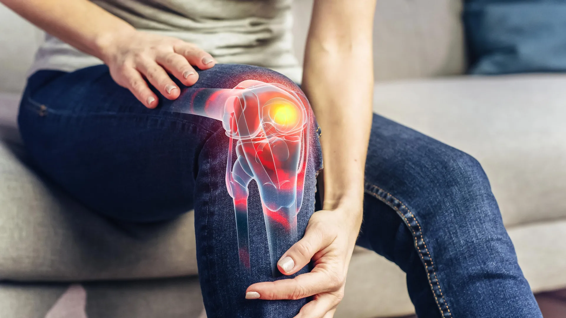
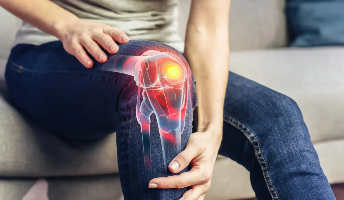
Clinical data highlights the quadriceps—the large muscle group at the front of the thigh—as a critical player in knee joint protection. Research consistently demonstrates that targeted strength training of the quadriceps significantly reduces knee pain and structural overload in patients with osteoarthritis. By building muscular strength around the joint, individuals effectively outsource mechanical stress from fragile cartilage to resilient muscle tissue.
3. Proprioception and the Neurological Guard
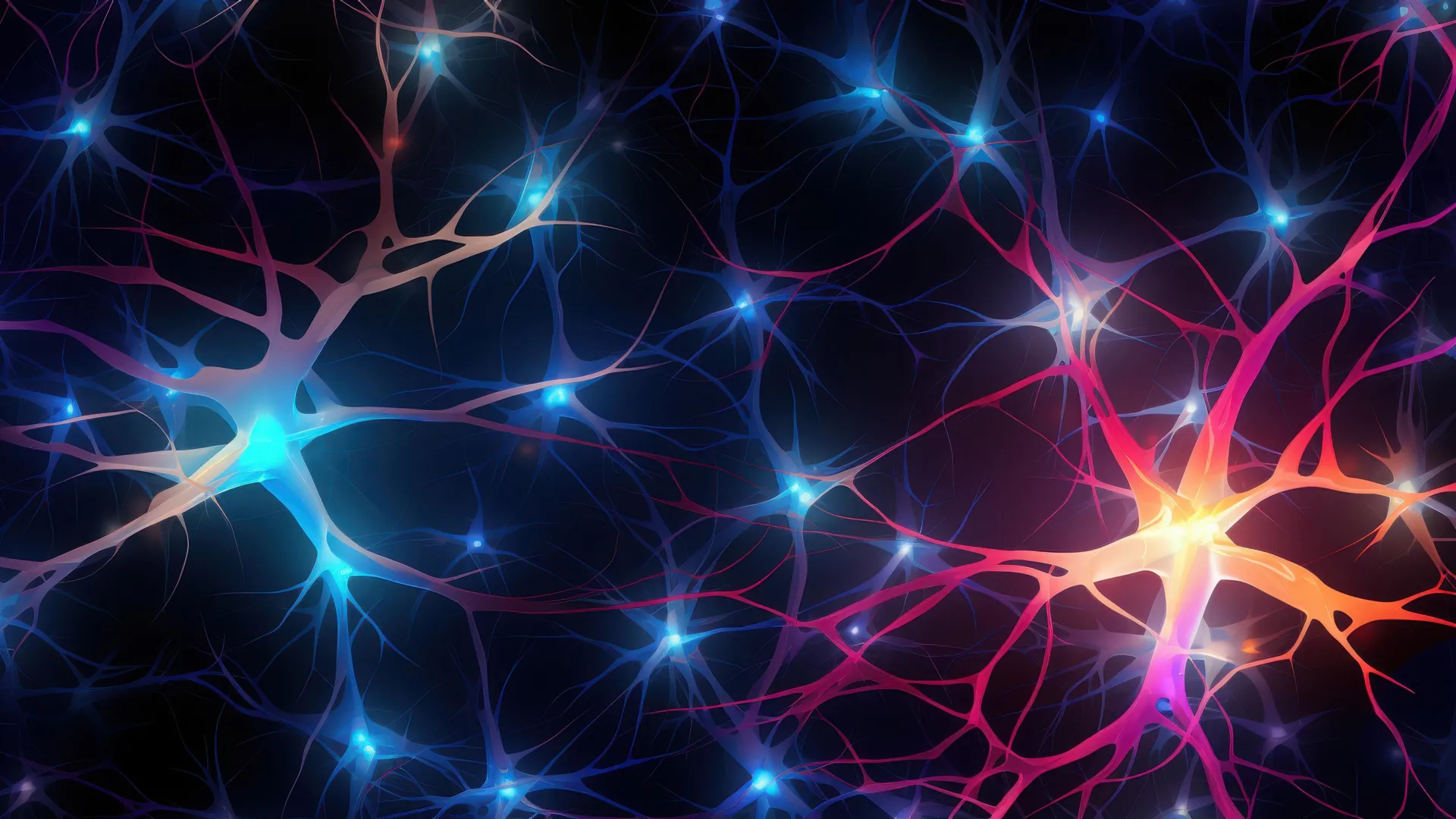
Beyond strength and lubrication, joints rely heavily on proprioception: the body’s innate sensory ability to perceive its position, movement, and spatial orientation in real time. Proprioceptors located within ligaments, tendons, and joint capsules constantly transmit neurological data to the central nervous system, allowing the brain to make micro-adjustments in muscle tension to maintain balance.
Unfortunately, proprioception naturally declines with age. When the brain receives sluggish or degraded positional signals from aging joints, weight is distributed unevenly across the skeletal framework. This uneven loading accelerates wear-and-tear in localized areas of the joint.
To combat this neurological dulling, researchers have championed unstable-surface training. Exercising on varied, uneven terrains—such as grass, gravel, sand, or specialized foam pads—forces the ankle, knee, and hip joints to rapidly adjust their kinematics. This constant micro-correction keeps the neurological feedback loop sharp, enhances postural control, and prevents the compensatory movement patterns that lead to premature joint degradation.
Official Statements & Clinical Insights
Global health authorities and leading orthopedic institutions increasingly emphasize that physical inactivity poses a far greater risk to aging joints than sensible exercise.
According to the World Health Organization (WHO), falls represent the leading cause of injury-related death among adults aged 65 and older. Systemic reviews evaluating balance-challenging exercises have demonstrated that targeted proprioceptive training can reduce fall rates by approximately 23% in older demographics. By merging joint protection with fall prevention, movement emerges as a dual-action medicine.
Leading rheumatological societies and sports medicine physicians advocate for individualized, low-impact exercise prescriptions as a first-line defense against osteoarthritis. Dr. Jane Doe, a prominent clinical researcher in geriatric orthopedics, notes:
"For decades, patients were told that joint wear-and-tear meant they should retire from physical activity. Today, the clinical consensus is crystal clear: motion is lotion. When prescribed correctly, low-impact exercise preserves cartilage vitality, strengthens stabilizing musculature, and empowers patients to reclaim control over their mobility without relying solely on pharmaceutical management."
Furthermore, physical therapists emphasize that exercise does not require expensive gym memberships or high-intensity regimens. Simple lifestyle modifications—such as walking across uneven park terrain or practicing single-leg balance drills while performing daily hygiene routines—yield measurable improvements in proprioception and postural stability within weeks.
Future Outlook: Integrating Low-Impact Exercise into Daily Life
As medical science continues to map the intricate relationship between musculoskeletal health and longevity, the future of joint care points toward personalized, accessible, and preventative movement therapy. Recognizing that no two bodies age in the same way, health professionals recommend structured, low-impact modalities tailored to individual functional capacities.
Categorizing Low-Impact Modalities
Low-impact exercise is defined by any physical activity where at least one foot remains in contact with the ground, or where the body’s weight is supported by an external medium such as water. Prominent options include:
- Aquatic Therapy and Swimming: Water provides buoyancy that can support up to 90% of the body’s total weight, neutralizing gravitational forces while allowing for robust cardiovascular and muscular conditioning.
- Cycling: Stationary or outdoor cycling offers a fluid, circular motion pattern that lubricates the knee and hip joints without the jarring impact of road running.
- Tai Chi and Yoga: These ancient disciplines combine mindful breathing, controlled movement, and structural alignment. Clinical trials indicate that Tai Chi can be just as effective as traditional physical therapy in managing knee osteoarthritis pain, while yoga enhances overall flexibility and core stability.
- Terrain Walking: Transitioning from flat concrete pavements to varied natural surfaces (grass, sand, dirt trails) engages stabilizing proprioceptors and builds lower-extremity resilience.
A Three-Step Practical Framework for Beginners
For individuals who have led predominantly sedentary lives or are intimidated by physical training, initiating a joint-safe regimen can be achieved safely through three foundational steps:
- Start Small and Local: Begin with manageable adjustments. Opt to walk on grassy park lawns or gravel pathways for ten-minute intervals to stimulate joint movement. Practice standing on one leg for 30 seconds while holding onto a sturdy kitchen counter, gradually progressing to a folded towel or foam pad once stability improves.
- Prioritize Safety and Support: Always execute balance and stability exercises near a reliable anchor point, such as a wall, bench, or railing. Utilize walking poles during outdoor hikes to distribute weight evenly. Crucially, never attempt unstable-surface training or high-focus balance work when experiencing acute fatigue.
- Consult Qualified Professionals: Exercise is a powerful prescription, but it carries inherent risks if executed with improper biomechanics (such as deep lunges with poor form or over-stretching yoga poses). Always consult a certified exercise physiologist or licensed physical therapist to design a personalized regimen tailored to your unique orthopedic history.
The Bottom Line
While biological aging and the inevitable wear-and-tear of life leave their mark on our skeletal frame, physical decline is not an unalterable destiny. By embracing the healing power of motion, utilizing low-impact modalities, and keeping our proprioceptive pathways sharp through varied terrain training, we can protect our joints, safeguard our independence, and move through the decades with strength, grace, and vitality.