









Subacute post-traumatic ascending myelopathy (SPAM) remains one of the most enigmatic, feared, and potentially devastating secondary complications following a traumatic spinal cord injury (SCI). Defined clinically as neurological deterioration involving the extension of deficits at least two spinal levels above the initial injury site within the first few weeks post-trauma—and occurring entirely in the absence of mechanical instability or syringomyelia—SPAM affects roughly 1% of all acute SCI patients. Because its pathophysiology is complex and multi-factorial, involving elements of vascular congestion, cytotoxic edema, inflammation, and altered cerebrospinal fluid (CSF) dynamics, it continues to challenge trauma clinicians worldwide.
To shed new light on this clinical enigma, a comprehensive case series and literature evaluation were recently conducted at the Hospital Universitari Vall d’Hebron (HUVH) in Barcelona, Spain. As a premier national trauma center and the designated reference hospital for acute traumatic spinal cord injuries across Catalonia—a region serving a population of approximately 7.9 million—HUVH is uniquely positioned to manage and analyze these rare occurrences. Over a seven-year span (2017–2023), investigators meticulously documented three distinct cases of SPAM out of 557 acute spinal cord injury admissions, yielding an institutional incidence rate of 0.54%.
This landmark retrospective analysis not only highlights the unpredictable nature of SPAM but also explores the comparative efficacy of differing corticosteroid regimens—specifically contrasting a descending dexamethasone taper with tailored intravenous methylprednisolone boluses. While the sample size underscores the sheer rarity of the condition, the insights gained from HUVH offer critical guidance for early diagnosis, advanced neuroimaging interpretation, and the refinement of acute neuro-intensive care protocols.
Detailed Chronology: The Vall d’Hebron Case Series
To understand the clinical trajectory of SPAM, one must examine the real-world presentation of affected individuals. The HUVH case series documents three distinct patients treated between 2017 and 2023, each presenting unique challenges in stabilization, delayed neurological decline, and pharmacological intervention.
Case 1: The Motorcyclist with Cervical Fracture-Dislocation
The first case involved a 41-year-old male with a medical history of type I diabetes who suffered severe polytrauma in a high-speed motorcycle accident. Upon arrival at the HUVH emergency department, the patient was hemodynamically stable. Advanced Body-CT imaging revealed a complex C6-C7 fracture with greater than 50% anterolisthesis, left facet dislocation, and a right facet fracture resulting in critical spinal canal invasion. Initial physical and neurological examinations established a C5 neurological level spinal cord injury classified as American Spinal Injury Association (ASIA) Impairment Scale (AIS) Grade A (complete motor and sensory loss), a finding corroborated by baseline cervical magnetic resonance imaging (MRI).
Forty-eight hours post-injury, the patient successfully underwent closed reduction and C6-C7 internal fixation and arthrodesis without acute surgical complications. However, the postoperative course proved turbulent. In the context of a descending dexamethasone taper, the patient struggled with poorly controlled blood glucose levels and hypertension. Concurrently, attempts to manage severe spasticity with baclofen triggered dangerous hypotensive episodes, necessitating a pharmacological switch to tizanidine.
Thirty-five days following the initial trauma, the clinical picture shifted dramatically. The patient reported acute, worsening neck pain accompanied by painful dysesthesias radiating across the neck and upper dorsal region, along with progressive paresthesias in both upper extremities. A focused neurological re-examination diagnosed a C3 spinal cord injury (AIS A)—representing a frightening two-level upward neurological regression.
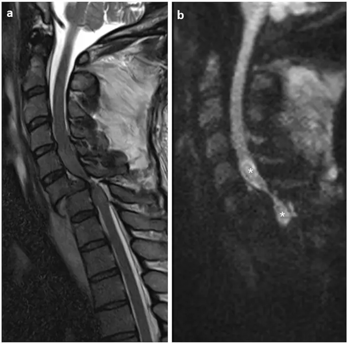
An urgent cervical MRI confirmed the diagnosis of SPAM, revealing a newly developed, extensive centromedullary edema ascending all the way to the C1 vertebral level. Clinicians initiated a secondary rescue course of intravenous dexamethasone (4 mg every 6 hours for three days). While this intervention halted further ascension and yielded a modest, one-level neurological improvement by the time of discharge, it failed to alter the patient’s baseline AIS classification.
Case 2: Polytrauma and Delayed Cervical Progression
The second case centered on a 45-year-old female smoker with underlying obesity who sustained critical injuries when struck by a vehicle. She arrived at the trauma center in a state of profound hemodynamic instability, requiring emergency intubation, aggressive volume resuscitation with blood products, and continuous vasoactive drug support to maintain adequate mean arterial pressure.
A comprehensive Body-CT cataloged an array of severe injuries: a C1 (Jefferson) fracture, a catastrophic burst fracture at the T2 level with significant spinal canal invasion, a basal right pneumothorax, bilateral hemothorax, right lung atelectasis, and a posterior mediastinal hematoma devoid of active bleeding. Furthermore, she suffered an open femoral fracture that necessitated immediate external fixation.
Following stabilization of her systemic physiology, cervico-dorsal MRI and physical assessments confirmed a C7 spinal cord injury (AIS A). Before spinal decompression could proceed, the patient developed diabetic ketoacidosis and required bilateral chest tube insertions to manage pulmonary complications. Forty-eight hours post-injury, surgeons performed a successful C6-T4 posterior instrumentation and T2-T3 fracture reduction.
The postoperative recovery was prolonged and fraught with respiratory challenges, requiring extended mechanical ventilation. She suffered a sudden hypoxic event characterized by desaturation and severe bradycardia driven by right basal atelectasis and an apical pneumothorax, which resolved following chest drain adjustments and non-invasive ventilatory support.
Thirty-nine days after the accident, the patient experienced a noticeable loss of function in both upper extremities. A clinical evaluation revealed a deterioration to a C4 spinal cord injury (AIS A)—a three-level upward migration of the spinal lesion. Urgent follow-up MRI demonstrated classical SPAM features, with centromedullary edema tracking upward to the C2 level, accompanied by signal heterogeneity and focal microhemorrhages. Medical staff administered a three-day course of intravenous methylprednisolone pulses (1 g/day). Although this high-dose steroid protocol failed to alter her overall neurological level or AIS grade by discharge, clinicians noted a measurable improvement in baseline muscle strength.
Case 3: Thoracic Burst Fracture and Ascending Conus Edema
The final case involved a 40-year-old male smoker with a history of type II diabetes who was transferred to HUVH following a traumatic workplace fall. Presenting with lower limb immobility, he was hemodynamically stable upon evaluation. Body-CT imaging revealed an unstable T8 burst fracture causing 30% to 40% spinal canal narrowing and severe anterior cord compression. Baseline assessments diagnosed a T7 spinal cord injury (AIS B), confirmed via thoracic MRI.
Surgical intervention was executed 48 hours post-trauma, consisting of a T7-T9 laminectomy for decompression coupled with T6-T10 posterior arthrodesis. Intraoperatively, the patient experienced an acute bronchospasm managed with targeted bronchodilators. The postoperative phase was complicated by massive left-lower-lobe lung atelectasis requiring therapeutic fibrobronchoscopy and patient repositioning, subsequent acute respiratory distress syndrome (ARDS) necessitating prone-position ventilation, and persistent glycemic instability. Extubation was achieved six days post-surgery.
Just eleven days following the initial injury, routine neurological monitoring detected an upward progression: the injury level had ascended to T5 (AIS A), marking a two-level neurological deterioration. Immediate dorsal MRI confirmed aggressive SPAM, with extensive edema stretching inferiorly from the conus medullaris up to the C7 level, alongside subacute ischemic and cytotoxic edema spanning from T2-T3 down to T6.
Unlike the previous two patients, Case 3 was treated with an extended, seven-day course of intravenous methylprednisolone pulses (1 g/day). The therapeutic response was markedly more favorable: upon discharge, the patient’s neurological status recovered to a T5 injury with an upgraded classification of AIS B, demonstrating partial sensory preservation below the neurological level and proving that targeted, prolonged high-dose corticosteroid therapy can alter the disease trajectory in select cases. Notably, across all three patients, no severe adverse systemic side effects were observed from the administered corticosteroid regimens.
Supporting Context, Metrics, and Pathophysiological Theories
To contextualize these clinical findings, the HUVH research team evaluated the broader epidemiological and mechanistic data surrounding SPAM. Affecting roughly 1% of all spinal cord injury patients globally, SPAM exhibits a pronounced demographic skew, predominantly striking young-to-middle-aged males with a reported male-to-female ratio reaching 5:1. In the HUVH cohort spanning 2017 to 2023, three out of 557 acute SCI admissions yielded an institutional incidence of 0.54%, aligning closely with international literature. The patients in this series averaged 42 years of age, fitting squarely within the high-risk demographic window.
Unraveling the Etiology
The precise pathophysiological mechanisms triggering SPAM remain intensely debated. While multiple hypotheses have been advanced, no single theory has achieved universal consensus. The primary suspected mechanisms include:
- Cerebrospinal Fluid (CSF) Dynamics: Alterations in normal CSF flow patterns and pressure gradients around the injury site are frequently cited as a primary catalyst for ascending cord edema.
- Vascular Compromise: Ischemia driven by arterial or venous thrombosis—such as occlusion of the Artery of Adamkiewicz, venous congestion, or prolonged hypotensive episodes—can precipitate secondary necrosis.
- Excitotoxicity and Apoptosis: Secondary biochemical injury cascades, cellular apoptosis, and localized inflammatory or autoimmune reactions can drive the progressive upward march of necrosis far beyond the boundaries of the primary mechanical impact.
- Systemic Physiological Stress: All three HUVH patients experienced notable systemic complications—including fluctuations in mean arterial pressure, severe pulmonary events (atelectasis, pneumothorax, ARDS), and glycemic dysregulation (diabetic ketoacidosis or poor diabetic control)—prior to or during the onset of SPAM. This clinical correlation underscores the hypothesis that systemic hypoxemia and vascular instability act as critical secondary triggers.
Diagnostic Benchmarks
Clinically, SPAM manifests as a progressive neurological decline occurring typically within the first three weeks post-injury (averaging 28.3 days in the HUVH series), with sensory and motor deficits extending an average of 2.3 levels above the primary lesion. Symptoms encompass severe neck, scapular, or trunk pain, ascending paresthesias, bimanual motor deficits, and, in severe cases involving the brainstem, respiratory failure or autonomic collapse.
Magnetic Resonance Imaging (MRI) remains the undisputed gold standard for diagnosis. Typical SPAM imaging features include extensive centromedullary T2-weighted hyperintensity spanning at least four vertebral segments above the injury epicentre, tapering gradually toward the rostral extent. Furthermore, diffusion-weighted imaging (DWI) and T2 sequences frequently reveal microhemorrhages and cytotoxic edema. However, clinicians must remain cognizant that radiological progression does not always mirror immediate clinical decline; edema can sometimes be visualized on MRI prior to overt clinical manifestation.
Official Statements and Clinical Perspectives
Reflecting on the management of these complex cases, the clinical team at the Hospital Universitari Vall d’Hebron emphasizes the critical need for heightened clinical vigilance in the weeks following acute spinal trauma.
"Subacute post-traumatic ascending myelopathy is an infrequent yet profoundly threatening complication that demands our utmost vigilance," noted the research group in their institutional review. "Because the window between initial stabilization and neurological deterioration is often narrow, continuous and systematic neurological mapping by specialized spinal cord injury units is absolutely vital for early detection."
Addressing the ongoing dilemma surrounding therapeutic protocols, the authors emphasized the limitations of current medical literature:
"Published literature provides no definitive consensus regarding the superiority of any single pharmacological or surgical intervention. While supportive care, surgical decompression, and various corticosteroid schedules remain the cornerstones of management, our limited case series suggests that prolonged intravenous methylprednisolone courses may be associated with more favorable clinical stabilization compared to dexamethasone tapers. Nonetheless, given the small patient cohorts typical of this rare condition, these observations must be interpreted with caution, and further multi-center trials are urgently required to establish an evidence-based standard of care."
Furthermore, the clinical staff highlighted the importance of aggressive multidisciplinary care:
"Our findings underscore that SPAM does not occur in a vacuum. The close intersection between systemic cardiovascular stability, strict glycemic control in diabetic patients, and pulmonary management highlights the reality that secondary spinal cord injury is deeply tied to whole-body homeostasis. Protecting the compromised spinal cord requires aggressive management of systemic blood pressure and prompt correction of pulmonary compromise."
Future Outlook and Clinical Recommendations
As the medical community continues to grapple with the unpredictable nature of SPAM, the insights compiled from the HUVH trauma registry point toward several vital pathways for future research and clinical protocol development:
- Standardized Surveillance Protocols: Given that SPAM typically strikes within the first three to five weeks following trauma, spinal injury centers should implement enhanced, high-frequency neurological and radiological monitoring protocols during this critical subacute window—especially for patients presenting with complete injuries at the thoracolumbar junction.
- Multicenter Collaborative Registries: Due to the low overall incidence of SPAM (~1%), single-center case series, while valuable, inherently lack the statistical power required to definitively prove the superiority of specific corticosteroid or surgical protocols. Establishing international, multicenter registries will be essential to aggregate sufficient data for robust prospective trials.
- Refining Pharmacological Therapies: Comparative studies are urgently needed to evaluate the pharmacodynamics of high-dose methylprednisolone versus alternative anti-inflammatory and neuroprotective agents, aiming to mitigate secondary apoptotic cascades without inducing systemic complications like hyperglycemia or immunosuppression.
- Holistic Intensive Care Integration: Future management guidelines must emphasize aggressive hemodynamic optimization and pulmonary care, recognizing that systemic insults (such as atelectasis, hypoxia, and blood pressure fluctuations) directly threaten vulnerable, recovering neural tissues.
Ultimately, while SPAM remains a formidable clinical challenge that frequently leaves patients with permanent neurological deficits, early recognition, rapid diagnostic imaging via MRI, and proactive multidisciplinary supportive care offer the best opportunity to halt its devastating upward progression and preserve remaining neurological function.