






Executive Overview
In the realm of rare hematology, the convergence of multiple complex pathologies presents an extraordinary diagnostic and therapeutic puzzle. A recent clinical case originating from the "Unidad Hemato-Oncológica Especializada" in Cúcuta, Norte de Santander, Colombia, has brought this reality to the forefront. Documented by a team of medical researchers, the case details the clinical trajectory of a 25-year-old male presenting with a rare dual diagnosis: Chediak–Higashi syndrome (CHS) complicated by an Inborn Error of Immunity-associated lymphoproliferative disorder (IEI-LPD), specifically featuring a T-cell lymphoma.
Chediak–Higashi syndrome is an exceedingly rare autosomal recessive condition characterized by lysosomal dysfunction, partial albinism, neurological impairment, and profound immunodeficiency. Globally, fewer than 500 cases have been formally documented in medical literature, making epidemiological tracking exceptionally difficult. When compounded with an aggressive inborn error of immunity-associated malignancy, the physiological barriers and treatment limitations multiply exponentially.
Despite initial therapeutic success using dose-adjusted chemotherapy regimens, the patient ultimately succumbed to progressive neurodegeneration and infectious complications. This case highlights the vital need for advanced genetic profiling, heightened clinical awareness, multidisciplinary management strategies, and the global imperative to bridge diagnostic disparities in regions burdened by socioeconomic and geopolitical strains.
Detailed Chronology: A Patient’s Journey
Presentation and Historical Context
The patient, a 25-year-old male, was referred to the hemato-oncology unit experiencing incapacitating lower back pain over the preceding week, coupled with a quantifiable weight loss of 5 kg (approximately 11 lbs) over a two-to-three-month window. These symptoms were accompanied by systemic complaints of asthenia, adynamia, severe fatigue, and notable skin pallor.
A review of his medical history revealed a significant prior intervention: left ocular surgery due to a suspected cataract, which was complicated by postoperative endophthalmitis and eventual complete loss of vision in the left eye.
Physical Examination and Diagnostic Workup
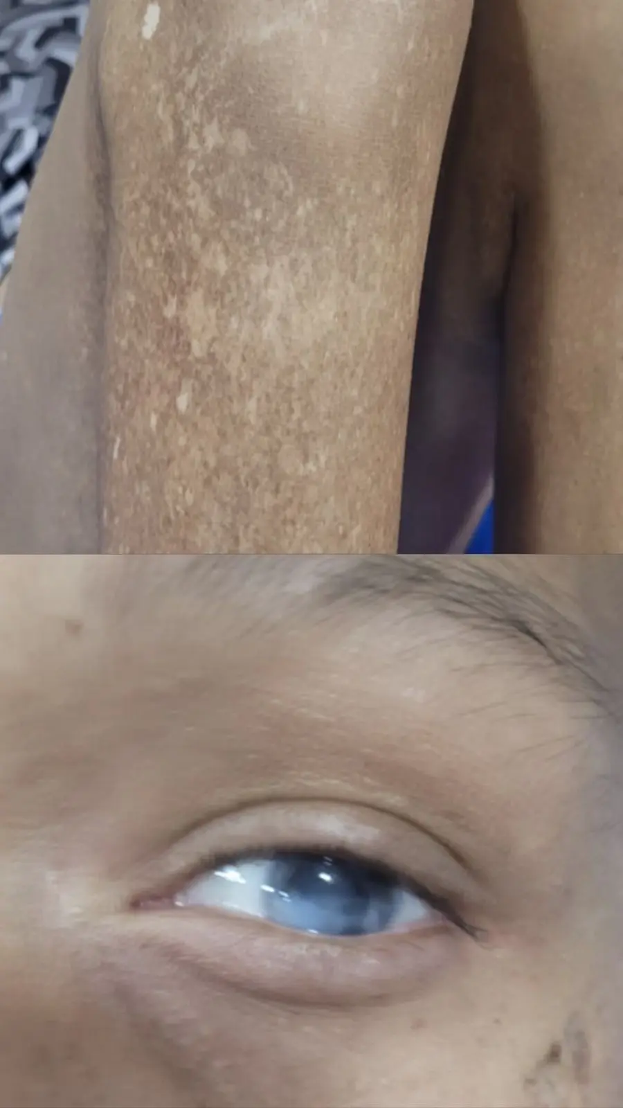
Upon clinical examination, the patient exhibited classic phenotypic markers of CHS:
- Dermatological and Integumentary Findings: Generalized skin pallor, multiple hypochromic maculae distributed across the body, and noticeable hair hypopigmentation.
- Ophthalmological Findings: Left ocular hypopigmentation located in the lower nasal quadrant, alongside a translucent left iris.
- Neuromuscular and Systemic Signs: Generalized hypotrophy (wasting) across the extremities and pronounced walking abnormalities.
Initial laboratory testing unveiled systemic compromise, including leukopenia (3,830 cells/µL), neutropenia (1,370 cells/µL), and moderate normocytic normochromic anemia with a hemoglobin level of 8.6 g/dL. A peripheral blood smear exposed striking abnormalities: hyperpigmentation alongside large, atypical cytoplasmic inclusions within both lymphocytes and neutrophils.
Serum protein electrophoresis confirmed hypogammaglobulinemia, characterized by depressed levels of IgA (19.4 mg/dL) and IgM (11.6 mg/dL), alongside a low total protein count of 4.7 g/dL and hypoalbuminemia (2.6 g/dL). Flow cytometry of the peripheral blood demonstrated an elevated count of CD3+ T lymphocytes, an inverted CD4/CD8 ratio (0.2), and a depleted natural killer (NK) cell population (73.2 cells/µL), confirming severe functional immune impairment.
Advanced Imaging and Histopathology
Magnetic resonance imaging (MRI) of the lumbosacral spine exposed compression fractures at the T12 and L1 vertebrae. More critically, imaging identified a large paravertebral retroaortic mass measuring $91 times 53 times 30text mm$ adjacent to the T10–T12 vertebral bodies, alongside concurrent splenomegaly. Bone scintigraphy confirmed blastic hyperactivity within the fractured T12 and L1 vertebrae.
A CT-guided Tru-cut biopsy of the retroaortic mass revealed a proliferation of small lymphoid-appearing cells arranged in sheets and loose nodules within a fibrous stroma. Morphological and immunophenotypic panels established a diagnosis of a primary immunodeficiency-associated T-cell lymphoproliferative disorder (T-cell IEI-LPD). Bone marrow aspiration reinforced these findings, showing intense granularity in the lymphoid and granulocytic series, medullary lymphocytosis, and a predominance of cytotoxic CD8+ T cells.
Genetic Sequencing and Molecular Insights
Genomic DNA extracted from peripheral blood samples was sequenced on an Illumina MiSeq platform. Bioinformatic evaluation identified a homozygous single-nucleotide variant within the LYST gene: c.9464G>A (p.Arg3155Gln), corresponding to dbSNP ID rs1266965849.
This non-synonymous substitution occurs in exon 40, specifically within the BEACH (Beige and Chediak-Higashi) domain of the 3,801 amino acid protein. Although traditionally classified in public databases as a variant of uncertain significance (VUS), its homozygous manifestation in familial presentations within regional populations strongly suggests a deleterious, pathogenic role driving the disease phenotype.
Supporting Context & Metrics
The Pathophysiology of Chediak–Higashi Syndrome
CHS is driven by mutations in the LYST (Lysosomal Trafficking Regulator) gene, located on chromosome 1q42.3. The LYST protein is crucial for the normal formation, storage, and secretory dynamics of lysosomal granules across various cell types, including leukocytes, fibroblasts, melanocytes, and platelet dense bodies.
Mutations disrupt these mechanisms, yielding abnormally enlarged, non-functional vesicles. In melanocytes, this failure prevents the proper transfer of melanosomes, resulting in the characteristic partial oculocutaneous albinism. In leukocytes, defective lysosomal exocytosis cripples antimicrobial defense and immune surveillance.
The Oncogenic Link: Inborn Errors of Immunity and Lymphoma
Patients with inborn errors of immunity (IEIs)—categorized under "Diseases of immune dysregulation" by the International Union of Immunological Societies—face a severely heightened risk of developing aggressive malignancies, notably lymphomas and lymphoproliferative disorders. This vulnerability stems from compromised T-cell immunosurveillance, defective DNA repair pathways, and chronic antigenic stimulation.
While lymphomas in IEIs commonly present with a bimodal age distribution and a male predominance, cases specifically overlapping with CHS are exceptionally rare, with fewer than 5,000 documented cases globally across medical literature. T-cell malignancies in this context are particularly aggressive and typically carry poor prognoses.
Treatment Protocol and Clinical Outcome
The patient was administered a therapeutic regimen consisting of six cycles of dose-adjusted EPOCH chemotherapy (etoposide, prednisone, vincristine, cyclophosphamide, and doxorubicin), paired with pegfilgrastim as primary prophylaxis against febrile neutropenia.
- Initial Response: Intermediate evaluations revealed a remarkable complete response, achieving 100% clearance of the retroaortic mass and normalization of spleen size.
- Complications and Decline: Institutional resource limitations precluded post-treatment Positron Emission Tomography (PET) scanning. Shortly thereafter, the patient experienced progressive neurological deterioration manifesting as a neurogenic bladder. This paved the way for secondary urinary tract infections, urosepsis, and ultimately, death.
- The Cytotoxic Dilemma: While vincristine is critical to the EPOCH regimen, its cumulative neurotoxic profile likely compounded the underlying neurodegenerative pathology inherent to CHS, illustrating the fine line oncologists walk between disease eradication and systemic toxicity.
Official Statements & Expert Perspectives
The reporting physicians and international hematology communities have emphasized several key takeaways from this complex case:
- On Diagnostic Delays: "Diagnosing rare conditions remains a profound challenge," noted the study’s corresponding author, Dr. Isabella Corona. Data indicates that a significant percentage of rare disease patients experience years of misdiagnosis. In this instance, historical misdiagnoses contributed to irreversible ocular complications prior to the hematological presentation.
- On Geographic and Socioeconomic Barriers: The clinical team candidly addressed the reality of practicing medicine in regions impacted by geopolitical instability and systemic resource limitations. Disruptions in healthcare infrastructure, medication supply chains, and advanced imaging modalities (such as PET scans) directly impact patient management and prognostic tracking in developing nations.
- On Genetic Classification: Regarding the LYST c.9464G>A variant, the authors advocate for its reclassification. Because the variant was detected in a homozygous state with a variant allele frequency (VAF) of 100%, and corroborated by familial presentations, the authors propose shifting its status from a Variant of Uncertain Significance (VUS) to a pathogenic mutation with probable deleterious functional outcomes.
Future Outlook
The convergence of Chediak–Higashi syndrome and T-cell IEI-LPD serves as a stark reminder of the frontiers remaining in rare disease research. Moving forward, the medical community must prioritize several strategic initiatives:
- Adoption of Next-Generation Sequencing (NGS): Expanding access to high-depth exome and genome sequencing will accelerate the identification of novel LYST variants, illuminating critical genotype-phenotype correlations.
- Standardization of Diagnostic Criteria: Unifying international terminology and establishing rapid diagnostic screening protocols for patients presenting with combined pigmentary and immunological anomalies can drastically reduce diagnostic latency.
- Multidisciplinary Management Frameworks: Given the multi-systemic nature of CHS—spanning neurology, immunology, dermatology, and oncology—care models must integrate cross-specialty oversight from early presentation through survivorship.
- Research into Curative Modalities: While intensive chemotherapy can induce temporary remissions in high-risk lymphomas, hematopoietic stem cell transplantation (HSCT) remains the only definitive curative approach for the underlying immunological and hematological defects of CHS, provided it is deployed before irreversible neurodegeneration sets in.
By documenting and analyzing cases of such profound rarity, the scientific community moves one step closer to demystifying the complex interplay between inherited immune dysregulation and oncogenesis, paving the way for targeted, personalized therapies that can alter the trajectory of otherwise fatal orphan diseases.