







Executive Overview
Sickle cell disease (SCD) remains one of the world’s most pervasive and challenging inherited blood disorders, characterized by severe hemolytic anemia, excruciating vaso-occlusive crises (VOCs), and progressive end-organ damage. At the cellular core of this pathology is the abnormal polymerization of deoxygenated sickle hemoglobin (HbS), which physically distorts red blood cells (RBCs) into rigid, fragile, crescent-like structures. These malformed cells lose their vital deformability, clumping together to obstruct microvascular blood flow.
Because mature red blood cells lack mitochondria, they depend entirely on glycolysis to generate intracellular adenosine triphosphate (ATP)—the essential energy currency required to maintain ion homeostasis, redox protection, membrane integrity, and cellular elasticity. In healthy individuals, RBCs also export controlled amounts of ATP to act as a crucial vasoregulatory signal, inhibiting intercellular adhesion and preventing capillary hyperpermeability.
Emerging therapeutic strategies aimed at altering RBC metabolism have centered on pyruvate kinase (PK) activators (PKAs), such as mitapivat (AG-348). By boosting the activity of red blood cell pyruvate kinase (PKR), these agents theoretically increase intracellular ATP production while simultaneously depleting upstream 2,3-diphosphoglycerate (2,3-DPG), an allosteric effector that otherwise promotes HbS deoxygenation and sickling.
However, a groundbreaking translational study led by researchers at Duke Health has revealed a surprising divergence in how healthy control (HC) and SCD red blood cells respond to these pharmacological agents ex vivo. While the PKA mitapivat successfully boosted intracellular ATP and ATP export in healthy control RBCs under both normoxic and hypoxic conditions, it failed to produce a statistically significant increase in intracellular or exported ATP within pediatric SCD patient samples. Intriguingly, however, exposing whole blood samples from SCD patients to the PKA did yield a significant anti-adhesive effect against laminin under physiologically relevant shear stresses, hinting at complex, multi-cellular mechanisms at play within whole blood environments.
Detailed Chronology and Experimental Methodology
To understand the physiological and biochemical boundaries of PKR activation in SCD, the research team designed a comprehensive ex vivo investigation comparing whole blood and isolated RBC samples from pediatric SCD outpatients against those of healthy adult volunteers.
Patient Cohorts and Sample Acquisition
The study evaluated whole blood samples obtained under institutional review board (IRB)-approved protocols. The SCD cohort comprised pediatric patients (ages 3 to 19 years) with confirmed homozygous SS genotypes, the vast majority of whom were actively undergoing hydroxyurea (HU) therapy. To ensure baseline integrity, patients who had experienced recent hospital admissions, blood transfusions, extreme fetal hemoglobin elevations, or treatment with specific alternative anti-sickling therapies within the prior 30 days were excluded.
The Ex Vivo Incubation Protocol
Whole blood samples were incubated with either 10 µM of the non-selective pyruvate kinase activator AG-348 (mitapivat) or a DMSO vehicle control. Initial exploratory runs on healthy control blood identified a standard incubation profile—18 to 24 hours at 4°C—that successfully elevated intra-RBC ATP and ATP export. This protocol was subsequently applied to the pediatric SCD cohort.
To determine whether temperature variations or shorter durations might unlock metabolic responsiveness in SCD samples, supplementary trials were conducted exposing cells to AG-348 for 1 hour at 4°C or 1 hour at 37°C. However, 24-hour incubations at 37°C were omitted after pilot testing revealed that prolonged physiological warming caused profound baseline ATP depletion in healthy control cells.
Tonometry, Hypoxia Exposure, and Luciferase Assays
Following incubation, RBCs were isolated, washed, and suspended in a Krebs buffer containing bovine serum albumin. Using a specialized rotating glass-bulb tonometer equilibrated to 37°C, the cells underwent controlled gas-mix exposure to simulate either normoxic (21% $O_2$) or hypoxic (1% $O_2$) microenvironments.
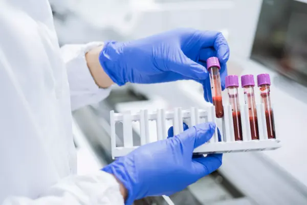
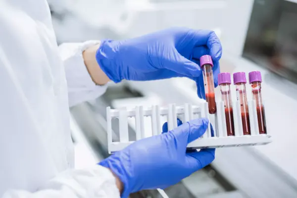
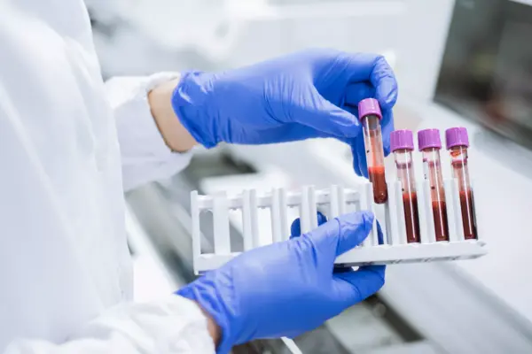
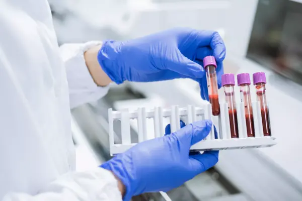
Following tonometry, supernatant concentrations (exported ATP) and intracellular ATP levels were quantified using a sensitive luciferase assay. Concurrently, spectroscopic hemoglobin analysis was deployed to calculate post-assay hemolysis rates, ensuring that any extracellular ATP detected was genuinely exported rather than leaked through cellular destruction. Cell lysis levels remained consistently low (averaging 1% to 2%) across all conditions, confirming that mechanical lysis did not skew the biochemical readouts.
Microfluidic Adhesion Assays
To evaluate functional outcomes, treated and control whole blood or isolated RBC suspensions were perfused through microfluidic channels pre-coated with recombinant human laminin (a major basement membrane substrate). Using a syringe pump, the channels were subjected to escalating physiological shear stresses ranging from 0.3 to 10 dynes/cm². Automated digital imaging captured cell retention rates at five-minute intervals, quantifying cellular adhesivity via image-processing software.
Supporting Context, Data, and Quantitative Metrics
The quantitative findings of the study illuminate a stark contrast between baseline metabolic profiles and drug responses in healthy versus sickle erythrocytes.
- Healthy Control Responses: Treatment of healthy control RBCs with AG-348 ex vivo confirmed prior literature, driving a robust, statistically significant rise in intra-RBC ATP during both normoxia (57.84 µM baseline vs. 77.59 µM post-AG-348) and hypoxia (57.67 µM baseline vs. 81.03 µM post-AG-348). Furthermore, untreated healthy control RBCs naturally escalated their ATP export when subjected to hypoxia (94.28 nM) compared to normoxia (79.70 nM). Exposing healthy control RBCs to AG-348 further amplified this export capacity during normoxia (109.0 nM) and hypoxia alike.
- SCD Patient Responses: In contrast, the pediatric SCD cohort demonstrated a muted metabolic response. Baseline intra-RBC ATP levels in the SCD cohort (nearly all on hydroxyurea) were comparable to healthy controls, defying the typical ~12% energy deficit historically observed in untreated adult SCD patients. However, ex vivo treatment with AG-348 failed to elicit a significant increase in intracellular ATP in SCD cells under normoxia (71.48 µM vehicle vs. 75.17 µM AG-348) or hypoxia. Similarly, exported ATP levels in SCD samples remained largely unchanged following drug exposure (79.70 nM vehicle vs. 77.12 nM AG-348 in normoxia).
- Adhesivity and Shear Dynamics: While treating isolated SCD red blood cells directly with AG-348 failed to alter their adherence to laminin, a striking shift occurred when whole blood was exposed to the drug. Whole blood exposure to AG-348 resulted in a statistically significant interaction ($p = 0.028$) across escalating physiological shear stresses (0.3, 1, and 3 dynes/cm²), demonstrating a clear reduction in SS RBC adhesion to laminin compared to the vehicle control. This discrepancy strongly implies that whole-blood elements—such as plasma proteins, leukocytes, or platelets—play a necessary modulatory role in mediating the anti-adhesive benefits of PK activation.
| Experimental Condition | Cohort | Exposure State | Intracellular ATP Change | Exported ATP Change | Hemolysis Rate |
|---|---|---|---|---|---|
| AG-348 vs. Vehicle | Healthy Control | Normoxia / Hypoxia | Significant Increase ($p < 0.01$) | Significant Increase ($p < 0.001$) | ~1% (No difference) |
| AG-348 vs. Vehicle | Sickle Cell Disease | Normoxia / Hypoxia | Non-significant ($p > 0.50$) | Non-significant ($p > 0.80$) | ~1% (No difference) |
| Whole Blood AG-348 | Sickle Cell Disease | Shear Stress (0.3–3 dyn/cm²) | N/A | N/A | Reduced Laminin Adhesion ($p = 0.028$) |
Official Statements and Author Insights
Lead authors and principal investigators emphasized the complex nature of translating acute ex vivo metabolic interventions into clinical paradigms for sickle cell disease.
"To our knowledge, our data show for the first time that ATP export from healthy control red blood cells increases after treatment with a pyruvate kinase activator—a process that simultaneously elevates intra-RBC ATP," noted Dr. Apoorva Jagadish, the study’s first author. "In contrast, exposure of SCD red blood cells to PKRA under identical conditions did not significantly increase intracellular or exported ATP during normoxia or hypoxia, even though whole-blood treatment successfully mitigated cellular adhesion."
Senior investigator Dr. Tim J. McMahon highlighted the clinical implications of these findings, particularly regarding the intersection of pharmacological therapy and existing treatment regimens:
"Most of our pediatric SCD cohort were actively maintained on hydroxyurea therapy, which has previously been associated with elevated baseline ATP production and release. It is plausible that these cells were already operating near an upregulated energetic ceiling, rendering acute ex vivo re-stimulation more difficult to achieve via single-dose PK activation alone. Moreover, the fact that whole-blood exposure—rather than isolated RBC exposure—reduced cellular adhesivity points toward critical cooperative signaling networks involving plasma or white blood cells that must be factored into future therapeutic evaluations."
Future Outlook and Clinical Implications
As pharmaceutical developers continue to advance red blood cell-specific pyruvate kinase activators (such as etavopivat and tebapivat) through clinical pipelines, this study provides vital cautionary insights and directions for future research.
- Investigating Long-Term Therapeutic Administration: Because acute ex vivo single-dose treatments fail to capture the cellular turnover, maturation cycles, and sustained metabolic shifts associated with chronic oral administration, future clinical trials must evaluate ATP export capacities directly from patients undergoing sustained, multi-week PKA therapy.
- Untangling Hydroxyurea Interplay: Researchers must determine whether baseline therapy with hydroxyurea uniquely alters the glycolytic reserve of pediatric and adult SCD erythrocytes, potentially masking the acute pharmacodynamic effects of secondary metabolic modulators.
- The Role of Whole-Blood Microenvironments: The demonstrated reduction in cellular adhesion following whole-blood PKA exposure—absent in washed isolated RBCs—demands deeper investigation into leukocyte-platelet-erythrocyte cross-talk. Understanding how extracellular ATP and purinergic signaling cascades modulate vascular inflammation will be paramount in preventing painful vaso-occlusive crises.
- Safety and Dosing Considerations: Because excessive extracellular ATP can occasionally act as a damage-associated molecular pattern (DAMP) that exacerbates acute inflammatory injury, the observation that SCD RBCs do not experience pathological surges in ATP export following PKA treatment offers reassuring safety data for the clinical translation of these emerging drugs.
Ultimately, while the metabolic mechanics of red blood cells in sickle cell disease present a labyrinth of regulatory checkpoints, refining our understanding of glycolytic modulation and intercellular signaling brings modern hematology one step closer to truly disease-modifying therapies.