








Executive Overview
In the rapidly evolving landscape of digital pathology, artificial intelligence (AI) has made tremendous strides in analyzing whole-slide images (WSIs). Yet, a fundamental gap has persisted between how AI models examine tissue slides and how human experts—veteran pathologists—actually navigate them. While standard vision-language models (VLMs) and computer vision architectures typically ingest static, high-magnification snapshots or process entire slides uniformly without contextual grounding, human experts utilize a dynamic, hypothesis-driven visual search strategy. They seamlessly transition between global structural overviews and microscopic, high-resolution region-of-interest (ROI) inspections.
A landmark study published in Nature Biomedical Engineering (July 2026), titled "Pathology-CoT: learning visual chain-of-thought agents from expert whole-slide image diagnosis behaviour," bridges this critical methodological divide. Spearheaded by Sheng Wang, Zhi Huang, and an interdisciplinary team of researchers primarily centered at the Perelman School of Medicine at the University of Pennsylvania, the study introduces Pathology-CoT—a framework that teaches vision-language agents to adopt the visual chain-of-thought and navigation behaviors of expert pathologists.
By capturing, parsing, and training AI agents on real-world pathologist viewport movements, the research team has demonstrated that models explicitly trained on human navigational logic significantly outperform standard proprietary and open-source models in complex diagnostic tasks. These tasks include identifying subtle micrometastases in colorectal cancer lymph nodes and classifying complex dermatopathological conditions.
This comprehensive review explores the methodology, validation processes, performance metrics, and broader clinical implications of the Pathology-CoT framework, drawing upon the study’s extensive supplementary and core data repositories.
Detailed Chronology and Methodological Architecture
The development of Pathology-CoT required the creation of an entirely new technical pipeline capable of translating erratic, high-frequency human viewing behaviors into structured, machine-interpretable visual reasoning steps.
1. Capturing and Preprocessing Pathologist Navigation
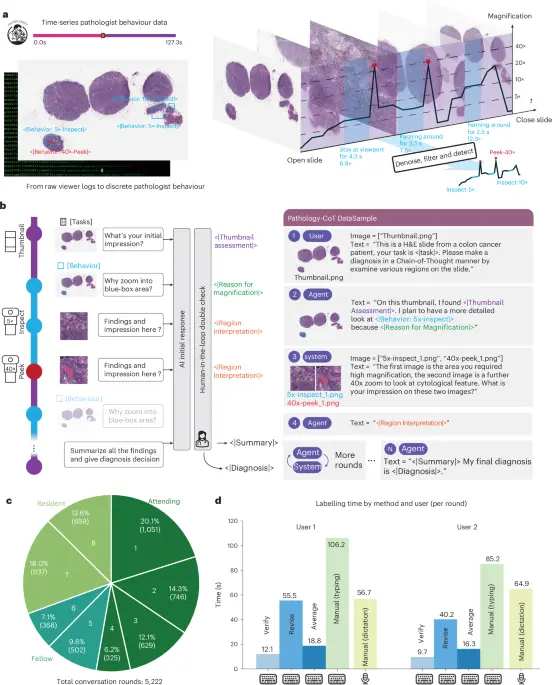
When a pathologist reviews a multi-gigabyte WSI, their interaction involves continuous zooming, panning, and lingering over specific cellular structures. To record this, the researchers deployed an automated AI Session Recorder that logs raw viewport dynamics—capturing mouse movements, zoom factors, and viewing windows in real time.
However, raw behavioral data is inherently noisy, plagued by overlapping viewports, redundant panning, and non-diagnostic overviews. To transform this raw telemetry into clean, actionable training data, the team engineered a sophisticated Behavior Analysis Pipeline (Extended Data Fig. 1):
- Raw Behaviour Ingestion: Initial high-frequency tracking data is captured as overlapping viewport coordinates.
- Initial Action Segmentation: Raw events are segmented using temporal heuristics into distinct behavioral categories, notably StayInspect (focused, stationary observation) and PanInspect (exploratory movement).
- Filtering Overview Actions: Excessively large, low-magnification bounding boxes corresponding to non-diagnostic slide-finding overviews are systematically eliminated.
- Merging Overlapping Actions: Spatially proximal actions displaying a high Intersection over Union (IoU) are consolidated into single, unified inspection events.
- Pruning Containing Actions: Redundant, larger bounding boxes that completely encapsulate smaller, highly specific inspection targets are pruned.
- Normalizing Action Bounding Boxes: The final action set is standardized to discrete magnification levels (e.g., $5times$, $10times$), generating a consistent, structured input format suitable for vision-language model consumption.
2. Validation Against Biological Eye-Tracking Data
To ensure that the AI Session Recorder accurately reflected true cognitive visual strategies rather than arbitrary mouse movements, the researchers validated the tool against biological eye-tracking data (Extended Data Fig. 2).
Comparative visualizations revealed stark differences between experience levels that the recorder successfully captured. First-year pathology residents exhibited highly exploratory, fragmented navigation patterns. In contrast, senior attending pathologists with decades of experience demonstrated highly targeted, efficient navigation. The discrete outputs generated by the AI Session Recorder mirrored ground-truth eye fixation points recorded by physical eye-trackers, confirming the reliability and biological fidelity of the captured data. Furthermore, tests across multiple independent sessions conducted by the same attending pathologist demonstrated remarkable consistency and stability in region-of-interest generation (Extended Data Fig. 3).
3. Training the Visual Chain-of-Thought Agent
With a reliable behavioral dataset established, the researchers trained specialized models—culminating in advanced variants such as Pathology-o3—to predict and execute expert-like visual reasoning chains. Rather than simply guessing a diagnosis from a static image tile, the model generates a sequential "thought process" mirrored by physical navigation: locating gross anatomical landmarks, identifying suspicious cellular anomalies, zooming into high-magnification details, and synthesizing clinical conclusions.
Supporting Context, Datasets, and Performance Metrics
The efficacy of the Pathology-CoT framework was rigorously evaluated across two major clinical cohorts: colorectal cancer (CRC) lymph node metastasis and complex skin cancer pathology.
The Diagnostic Cohorts
- Stanford CRC Lymph Node Cohort (LNCO2): Focused on identifying metastatic lesions within lymph nodes, a notoriously challenging task where small clusters of cancer cells can be easily overlooked. Whole-slide images and expert annotations from the LNCO2 dataset were acquired via the AIDA Data Hub.
- DROID Skin Cancer Cohort: Focused on dermatopathology, encompassing a wide array of neoplastic and inflammatory skin conditions. WSIs and annotations were likewise secured through the AIDA Data Hub.
Stratified Performance on Tumor Burden
One of the study’s most striking findings relates to performance stratification based on tumor burden in colorectal cancer lymph node metastasis (Extended Data Fig. 4a). Cases were categorized by difficulty:
- Isolated Tumor Cells (ITCs): $le 0.2text mm$ ($n=5$)
- Micrometastases: $0.2text–2text mm$ ($n=13$)
- Macrometastases: $>2text mm$ ($n=29$)
When benchmarked against leading proprietary models (such as GPT-4.1) and advanced open-source architectures (such as InternVL3 and Llama-4), Pathology-o3 consistently demonstrated superior localization and diagnostic accuracy, particularly in managing high-difficulty ITCs and micrometastases.
Crucially, qualitative visualizations of zoom-in behaviors demonstrated that Pathology-o3 mimics the exact spatial attention patterns of human attending pathologists (Extended Data Fig. 4b). When evaluating micrometastases and ITCs—which often lack the characteristic desmoplastic stromal reaction found in larger tumors—generic models frequently drifted toward irrelevant slide artifacts. In contrast, Pathology-o3 successfully targeted and inspected the precise micro-anatomic zones required for accurate staging.
Quantitative Attribution of Attention Drivers
Using the DROID segmentation ontology, the researchers conducted a quantitative attribution analysis to understand what drives the model’s visual attention (Extended Data Fig. 5).
Evaluating 960 high-confidence regions of interest predicted by a YOLOv8-Large architecture revealed that approximately 62.9% of model attention was allocated to normal anatomical structures (such as healthy epidermis and skin appendages) rather than explicit pathology. This reflects a genuine "screening" behavioral pattern akin to human workflow, where experts first establish baseline normal architecture before homing in on abnormalities.
Among the ROIs classified explicitly as "Pathology" ($N=340$), the model identified not only frank neoplasms (basal cell carcinoma, squamous cell carcinoma, and melanocytic neoplasms) but also subtle non-neoplastic findings—such as inflammation and cellular scarring—accounting for 33.5% of pathological foci.
Ablation Studies on Model Scale
An ablation study investigating YOLOv8 architectures of varying parameter scales (ranging from Nano to Large) on dermatopathology ROI prediction yielded vital engineering insights (Extended Data Fig. 7). While smaller models (Nano/Small) were frequently misled by local artifacts, incorrectly flagging staining ink or microscopic blood pooling as regions of interest, larger models (Medium/Large) exhibited a robust understanding of global tissue structure. They successfully prioritized anatomical context while ignoring visual distractors.
Official Statements and Institutional Contributions
The research represents a collaborative achievement between computational scientists, computer vision experts, and practicing clinical pathologists.
- Corresponding Author Attribution: The study was supervised by Dr. Zhi Huang of the Perelman School of Medicine at the University of Pennsylvania, who provided critical startup funding and oversight for the initiative. Correspondence regarding the study can be directed to
[email protected]. - Core Development Team: Methodology and experimentation were driven by primary contributors S. Wang, R. Wu, S. Liu, and Y. Liu, with data curation supported by J. Sun.
- Pathological Expertise: Vital expert pathology annotations, qualitative feedback, and clinical ground-truth validation were contributed by C. Herndon, S. Khosravi, X. Xu, D. E. Elder, J. A. Mills, A. Jukic, I. Hülsmann, M. Datta, and J. Saltz.
- Peer Review Acknowledgments: The editors of Nature Biomedical Engineering formally acknowledged the rigorous peer-review contributions of external experts, including Narayan Hegde and Hamid R. Tizhoosh, whose insights helped refine the evaluation benchmarks.
Future Outlook and Clinical Translation
The introduction of Pathology-CoT marks a philosophical shift in medical artificial intelligence. For years, the prevailing paradigm in computational pathology has relied on "black box" end-to-end classification models that map pixels directly to diagnoses without offering insight into how the conclusion was reached. This lack of transparency has historically hindered clinical adoption, as pathologists are legally and ethically bound to verify diagnostic rationales.
By aligning AI reasoning with human navigational telemetry, Pathology-CoT achieves two vital milestones:
- Interpretable AI Workflows: Clinicians can inspect the AI’s "visual chain-of-thought"—reviewing the exact progression of zoom levels, pan coordinates, and region-of-interest selections that led to a diagnostic recommendation.
- Enhanced Educational Utility: The recorded navigation patterns of master pathologists can be leveraged to train resident physicians, visualizing expert visual search strategies side-by-side with trainee behaviors.
Data and Code Availability
To foster open science and accelerate downstream research, the research team has made all primary resources publicly available:
- Source Code & Dataset: The Pathology-CoT Chain-of-Thought dataset for the skin cancer cohort, along with trained behavior predictors for both the Stanford CRC lymph node and skin cancer cohorts, is accessible via GitHub at github.com/zhihuanglab/Pathology-CoT.
- Whole-Slide Imaging Repositories: Raw WSIs and expert annotations from the LNCO2 and DROID skin datasets are available via the AIDA Data Hub upon application and formal institutional approval (LNCO2 Hub; DROID Hub).
As multimodal AI models continue to integrate into hospital information systems, frameworks like Pathology-CoT pave the way for transparent, highly accurate, and truly collaborative diagnostic assistants that work alongside pathologists as digital partners rather than opaque competitors.