









Executive Overview
In a monumental development for oncology and materials science, a collaborative research team from Weill Cornell Medicine and the Cornell Duffield College of Engineering has engineered a novel class of ultrasmall silica nanoparticles capable of executing a multi-pronged assault on aggressive prostate cancer. Published in the June 15 issue of the prestigious journal Cancer Research—a flagship publication of the American Association for Cancer Research—the preclinical study reveals that these engineered particles not only directly dismantle tumor cells but also dynamically reprogram the body’s immune defenses.
By flipping the biological switch of the tumor microenvironment from an immune-resistant "cold" state into a hyper-responsive "hot" state, the targeted nanoparticles have achieved unprecedented complete tumor remissions in murine models of aggressive prostate cancer. This dual-action paradigm bypasses traditional limitations of single-target therapies, simultaneously driving cancer cells toward a specialized form of self-destruction while breaking down metabolic and immune barriers that typically shield tumors from destruction.
Led by Dr. Michelle Bradbury and Dr. Ulrich Wiesner, the research represents the culmination of a long-running, cross-disciplinary collaboration bridging advanced materials engineering with cutting-edge molecular imaging and oncological therapeutics. As the investigative team prepares to transition these findings toward human clinical trials, the medical community is taking note. If successfully translated, this biocompatible, silica-based technology could radically redefine the treatment landscape for advanced, treatment-refractory prostate cancers, unlocking the full, long-dormant potential of modern immunotherapies.
Detailed Chronology: The Evolution of C’ Dots from Imaging Agents to Cancer Killers
The journey from diagnostic imaging tools to potent cancer therapeutics is a testament to the serendipitous and iterative nature of translational research. The foundational technology behind this breakthrough began years ago in the laboratories of Dr. Bradbury and Dr. Wiesner, focusing on diagnostic precision rather than direct cytotoxicity.
Origins as Diagnostic Tools: The Birth of C’ Dots
Originally developed to enhance the clarity and resolution of medical imaging during image-guided surgeries, the nanoparticles—formally designated as ultrasmall fluorescent core-shell silica nanoparticles, or Cornell Prime dots (C’ dots)—were engineered with exceptional precision. Composed of amorphous silica, a naturally occurring form of silicon dioxide found abundantly in foods, leafy greens, cereal grains, and the fossilized remains of microscopic organisms, these particles exhibited stellar biocompatibility.
Because of their sub-10-nanometer dimensions, uniform structure, and bright fluorescent cores, C’ dots excelled at navigating the human circulatory system, homing in on pathological tissues, and providing surgeons with real-time molecular maps of cancerous lesions. These diagnostic formulations have already cleared rigorous safety hurdles, successfully advancing into late-stage human clinical trials for image-guided surgical navigation and therapeutic delivery.
The Pivot Toward Therapeutics
However, a profound biological discovery altered the trajectory of the research. While studying the interaction of C’ dots with malignant cells, the investigators observed that the particles were not merely passive beacons; under specific configurations, they began to selectively damage cancer cells while leaving surrounding healthy tissues largely unharmed.
Recognizing the immense therapeutic potential of this phenomenon, the research teams launched a deep mechanistic investigation. Led by co-first authors Dr. Nabil Siddiqui, Dr. Li Zhang, and Dr. Gabriel DeLeon, alongside graduate students Nada Naguib and Rachel Lee, the team began engineering specialized targeting mechanisms onto the silica framework. They attached a high-affinity targeting molecule designed to explicitly recognize and bind to prostate-specific membrane antigen (PSMA), a well-established surface biomarker heavily overexpressed on the membranes of prostate tumor cells.
Preclinical Validation in Aggressive Prostate Cancer Models
To test the efficacy of these targeted particles, the team deployed advanced mouse models exhibiting aggressive, treatment-resistant prostate cancer. The administration of the PSMA-targeted C’ dots yielded extraordinary biological shifts. Not only did the particles home directly to the tumor sites with minimal off-target accumulation, but they also initiated a cascade of intracellular and immunological events that fundamentally altered the fate of the disease.
Mice treated with the optimized nanoparticle formulations showed striking reductions in tumor volume. Crucially, when these silica nanoparticles were integrated into multi-modal combination therapies involving immune checkpoint blockade and macrophage-targeting agents, the survival curves shifted dramatically. The treatment induced complete and near-complete remissions in a significant fraction of the test subjects, sparking intensive planning for clinical translation.
Supporting Context & Metrics: Mechanisms of Action and Data Breakdown
Understanding how an inert, naturally occurring substance like amorphous silica can orchestrate the destruction of malignant tissue requires an examination of the cellular and metabolic mechanics at play. The Weill Cornell study highlights two primary mechanisms: the induction of ferroptosis and the large-scale remodeling of the tumor microenvironment.
Triggering Ferroptosis: The Iron-Driven Oxidation Cascade
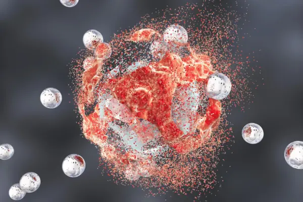
One of the most scientifically riveting findings of the study involves the induction of "ferroptosis"—a specialized, non-apoptotic form of programmed cell death driven by overwhelming, iron-dependent intracellular oxidation.
Unlike apoptosis (classic programmed cell death), which cancer cells frequently manage to evade through genetic mutations, ferroptosis operates via a fundamentally different biophysical route:
- Lipid Peroxidation: Intracellular iron ions catalyze chemical reactions that generate massive amounts of reactive oxygen species (ROS). These oxygen radicals specifically target and degrade polyunsaturated fatty acids embedded within the cell membranes.
- Membrane Collapse: As the lipid molecules of the membrane undergo cascading oxidation, structural integrity fails entirely, causing the cell to rupture and die from the inside out.
- Iron Transport: Investigators discovered that the silica nanoparticles—initially designed to carry diagnostic imaging agents—act as molecular shuttles. They scavenge and collect positively charged iron ions circulating within the bloodstream, transporting them directly across the cell membrane into the interior of the tumor cell.
Once concentrated inside the malignant cell, these imported iron ions act as the primary fuel source for the intense oxidative stress that drives ferroptosis, systematically dismantling the cancer cell’s structural defenses.
[Silica Nanoparticle (C' dot)]
       │
       ├──> Binds to PSMA Surface Receptor on Prostate Cancer Cell
       │
       ├──> Transports Circulating Iron Ions (Fe²⁺/Fe³⁺) into Cell Interior
       │
       ├──> Catalyzes Massive Intracellular Reactive Oxygen Species (ROS)
       │
       ├──> Drives Lipid Peroxidation of Cell Membranes
       │
       └──> Results in Structural Collapse & Cell Death via Ferroptosis
Transforming the Tumor Microenvironment (TME)
Beyond directly killing individual tumor cells, the nanoparticles exert a profound systemic effect on the surrounding cellular ecosystem, known as the tumor microenvironment (TME).
Solid tumors frequently establish an immunosuppressive "cold" microenvironment, characterized by the depletion of active T cells, the presence of immunosuppressive regulatory cells, and metabolic roadblocks that prevent the immune system from recognizing the cancer as a foreign threat. The Weill Cornell study demonstrated that the administration of C’ dots completely disrupted this protective shield:
- Immune Awakening: Inactive or exhausted T cells, tumor-associated macrophages, and antigen-presenting cells located within the TME were rapidly shifted into an active, aggressive, anti-tumor state.
- Metabolic Disruption: The nanoparticles disrupted core metabolic pathways across multiple cell types within the tumor niche, starving the cancer cells of essential nutrients and restricting their proliferative capacity.
- Sensitizing to Immunotherapy: By converting the "cold" tumor into an inflamed, immune-active "hot" tumor, the nanoparticles made the cancer remarkably vulnerable to standard immunotherapy drugs that had previously proven ineffective against prostate malignancies.
Breakdown of Combination Therapy Efficacy Metrics
The survival studies conducted on mice bearing aggressive prostate cancer yielded quantifiable metrics that underscore the synergy of multi-modal treatment regimens:
| Treatment Regimen | Observed Biological Effect | Complete Remission Rate |
|---|---|---|
| Control (No Treatment) | Rapid tumor progression, aggressive proliferation | 0% (Zero survival past baseline window) |
| C’ Dots Monotherapy | Modest survival improvement, localized cell death | Minimal / Low |
| Immune Checkpoint Blockade Alone | Standard checkpoint inhibition; limited efficacy in "cold" tumors | Minimal / Low |
| C’ Dots + Immune Checkpoint Blockade | Synergistic immune activation and direct cell killing | 40% (4 out of 10 mice achieved complete remission & indefinite survival) |
| C’ Dots + Checkpoint Blockade + CSF-1R Blockade | Maximum therapeutic synergy (targeting tumor-associated macrophages) | 50% (5 out of 10 mice achieved complete remission & indefinite survival) |
Safety evaluations accompanying these efficacy metrics revealed an exceptionally favorable profile. Although minor, transient accumulation of the particles was observed in peripheral filtration organs such as the spleen, histological and biochemical analyses showed no signs of systemic toxicity, organ damage, or adverse side effects in healthy tissues.
Official Statements & Expert Perspectives
The breakthrough has generated immense excitement across the academic and clinical medical communities, drawing praise from leading oncologists, radiologists, and materials scientists involved in the project.
Dr. Michelle Bradbury, the Endowed Professor of Imaging Research in Radiology, director of the Molecular Imaging Innovations Institute at Weill Cornell Medicine, and neuroradiologist at NewYork-Presbyterian/Weill Cornell Medical Center, emphasized the paradigm-shifting nature of the research:
"We’re very encouraged by these results; a treatment that directly induces tumor-cell death while transforming the immune microenvironment, as this does, would represent a new clinical paradigm. It is rare to observe a therapeutic agent that can orchestrate direct cytotoxicity while simultaneously rewriting the rules of the immune landscape."
Dr. Ulrich Wiesner, the Spencer T. Olin Professor in the Department of Materials Science and Engineering at Cornell University and co-corresponding author, marveled at the biological harmony displayed by the silica matrix:
"It seems unreal—how is it possible that rather than a single pathway we see all these effects happening simultaneously and only in tumors and not in healthy tissues? I have to wonder whether ultrasmall silica’s very early and ubiquitous presence in the environment and foods like leafy greens or cereal grains has given it a connection to biology that we’re only beginning to glimpse."
Adding clinical context to the findings, Dr. Jedd Wolchok, Meyer Director of the Sandra and Edward Meyer Cancer Center, professor of medicine at Weill Cornell Medicine, and director of the Parker Institute for Cancer Immunotherapy at Weill Cornell Medicine, highlighted the implications for immunotherapy resistance:
"One of the most intriguing aspects of this work is the convergence of direct tumor cell killing with broad immune remodeling. By creating conditions that support a more effective antitumor immune response, these particles may help unlock the full potential of immunotherapy in prostate cancer, where durable responses have historically been difficult to achieve."
The collaborative nature of the study was further underscored by Dr. Bradbury, who commended the tireless contributions of the research personnel, including co-first authors Drs. Nabil Siddiqui, Li Zhang, and Gabriel DeLeon, alongside graduate student contributors Nada Naguib and Rachel Lee, whose rigorous synthesis and characterization of the nanoparticles anchored the entire experimental framework.
Future Outlook & Clinical Translation
As the Weill Cornell and Cornell Duffield College of Engineering research consortium looks toward the horizon, the primary strategic objective is clear: bridging the gap between preclinical murine models and human clinical trials.
Charting the Path to Clinical Trials
Translating the success seen in animal models into human patients requires a structured, multi-step regulatory and clinical roadmap:
- Advanced Pharmacokinetics and Toxicology: Conducting exhaustive, large-animal safety studies to map the clearance, biodistribution, and long-term biocompatibility of the PSMA-targeted C’ dots in non-rodent mammalian models.
- Manufacturing Standardization: Scaling up the precise chemical synthesis of the core-shell silica nanoparticles under strict Current Good Manufacturing Practice (cGMP) guidelines to ensure absolute batch-to-batch uniformity, stability, and sterility.
- Phase I Human Clinical Trials: Designing early-phase clinical protocols focused on patients with advanced, metastatic, treatment-refractory prostate cancer. These initial trials will primarily evaluate human safety profiles, determine optimal dosing thresholds, and monitor preliminary signs of anti-tumor activity.
- Combination Therapy Trials: Subsequent trial phases will test the tripartite combination strategy—pairing the silica nanoparticles with immune checkpoint inhibitors and macrophage-modulating agents (such as CSF-1R blockers)—to replicate the high-rate complete remissions observed in the preclinical data.
Broader Implications for Oncology
Beyond prostate cancer, the foundational mechanics of this technology open up sweeping new vistas for cancer therapeutics. Because the underlying design relies on targeted delivery, iron scavenging, and the induction of ferroptosis, the platform could theoretically be adapted to target a wide array of solid tumors by swapping out surface biomarkers. By engineering different targeting peptides onto the outer silica shell, researchers hope to apply this dual-action strategy to breast, lung, pancreatic, and brain cancers that currently resist conventional therapies.
In an era where cancer treatment often relies on adding multiple high-toxicity drugs together, the prospect of utilizing an ultrasmall, naturally derived silica nanoparticle to simultaneously destroy tumor cells, starve their metabolism, and awaken the immune system offers a refreshing and profoundly hopeful horizon. If human trials mirror the success of these preclinical milestones, medicine may soon possess a powerful new weapon capable of turning the tide against the most aggressive malignancies known to humankind.