






Executive Overview
For decades, evolutionary biologists have mapped the branching tree of life with a sense of linear inevitability, assuming that complex anatomical structures evolve along steady, progressive trajectories. However, groundbreaking new research from Lund University and the University of Sussex shatters this conventional paradigm. According to a comprehensive comparative analysis of light-detecting cellular structures across the animal kingdom, humans and every other living vertebrate share a deeply improbable, almost mythological ancestor: a tiny, worm-like marine organism that navigated the primordial oceans roughly 600 million years ago with a single, central eye positioned squarely on top of its head.
This "cyclops-like" phase was not merely a quirky evolutionary footnote. Rather, it represents a critical pivot point in anatomical history. The new findings propose that our distant ancestors initially possessed paired eyes—standard equipment for mobile organisms navigating a complex three-dimensional environment. Yet, as these creatures transitioned to a sedentary, filter-feeding lifestyle, those paired visual organs atrophied and vanished through disuse. In their place, a solitary median eye persisted, serving as a primitive compass to distinguish night from day and orient the organism within the water column.
Millions of years later, when these ancient animals abandoned their stationary habits to return to an active, swimming lifestyle, evolutionary pressures demanded a sophisticated new visual apparatus. Instead of reinventing sight from scratch, nature pulled off an anatomical masterclass in recycling: it repurposed components of the ancient median eye to construct an entirely new pair of image-forming eyes. This tortuous, roundabout evolutionary detour explains a long-standing biological mystery that has puzzled scientists for generations: why vertebrate eyes—including our own—are fundamentally constructed in a manner diametrically opposed to the eyes of invertebrates like insects and cephalopods.
Furthermore, the study reveals that this ancient median eye never truly vanished from the biological blueprint. Its cellular and physiological ghost lingers deep within the modern vertebrate brain as the pineal gland, the endocrine master-regulator that governs our circadian rhythms and sleep cycles. By bridging comparative zoology, neurobiology, and evolutionary genetics, this research forces a radical reassessment of how the human brain and visual system came to be, proving that our waking and sleeping lives are inextricably bound to the singular gaze of an ancient ocean-dwelling ancestor.
Detailed Chronology: The 600-Million-Year Evolution of Vertebrate Vision
To trace the lineage of human sight, paleontologists and evolutionary biologists must look far beyond the emergence of the first fish, well before the colonization of land, and deep into the Ediacaran period. The narrative of vertebrate vision unfolds across four distinct evolutionary chapters, marked by radical environmental shifts and astonishing physiological adaptations.
Phase I: The Primal Dual-Eye Blueprint
Long before the divergence of the vertebrate lineage, early Precambrian organisms possessed rudimentary visual systems. In the earliest iterations of our distant forebears, this meant having two distinct groups of light-sensitive cells positioned on either side of the head. In evolutionary biology, paired eyes are the default adaptation for mobile animals. By comparing input from two separate points, a moving organism can calculate depth, judge distances, detect the trajectory of approaching predators, and orient itself within a spatial grid.
For these early marine denizens, bilateral symmetry and paired visual spots worked in tandem. However, the capricious pressures of natural selection do not favor complexity for its own sake. When maintaining a complex trait costs metabolic energy without offering a survival advantage, evolution ruthlessly trims the fat.
Phase II: The Sedentary Retreat and the Cyclopean Bottleneck
Approximately 600 million years ago, a pivotal behavioral shift occurred within our ancestral line. A group of small, worm-like marine creatures abandoned their active swimming habits, anchoring themselves to the ocean floor to adopt a stationary, filter-feeding existence. Suspended in the ocean currents, these organisms spent their lives passively sweeping the water for microscopic plankton.
In this low-stakes ecological niche, the active navigation tools of their ancestors became redundant. Complex, paired image-forming eyes required significant neural processing power and cellular maintenance, yet offered virtually no survival utility to a creature that no longer moved. Over countless generations, natural selection systematically dismantled the paired visual system. The lateral eyes atrophied and disappeared entirely from the physical form.
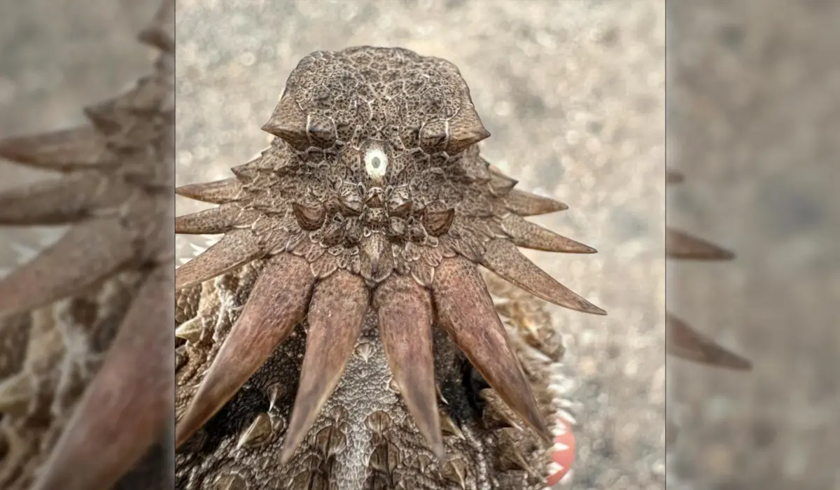
Yet, complete visual blindness was a liability, even for a stationary filter-feeder. The organism retained a small, centralized cluster of photoreceptive cells situated on the crown of its head. This solitary median eye—resembling the mythical cyclopean gaze—was sufficient for the creature’s basic ecological needs. It could not perceive sharp shapes or process high-resolution images, but it excelled at two crucial tasks: detecting the ambient shift from day to night (circadian entrainment) and discerning the upward direction of sunlight piercing the water’s surface (phototaxis). This single-eyed phase served as a genetic and physiological bottleneck through which all future vertebrates had to pass.
Phase III: The Great Return to Mobility and the Invention of the Vertebrate Retina
As epochs rolled forward, environmental pressures shifted once more. Descendants of these stationary worm-like creatures began to detach from the ocean floor, returning to an active, mobile lifestyle as swimming predators and foragers. Navigating a complex marine ecosystem demanded a renewed suite of sensory organs. To hunt prey, avoid hazards, and master dynamic surroundings, these animals desperately needed advanced vision.
Faced with this evolutionary crisis, nature executed a remarkable structural pivot. Rather than re-evolving lateral eyes from scratch or adopting the developmental pathways utilized by other successful phyla, evolution co-opted the existing median eye structure. Portions of this central visual apparatus were duplicated, expanded, and structurally transformed to generate an entirely new pair of image-forming eyes.
This unorthodox genesis explains one of the most profound structural dichotomies in zoology. In insects (such as fruit flies and bees) and mollusks (such as squid and octopuses), the retina develops directly from the surface ectoderm—the embryonic tissue that forms the outer skin of the head. In stark contrast, the vertebrate retina develops as an direct outgrowth of the embryonic brain itself. When light enters our eyes, it passes through layers of neural tissue before hitting the photoreceptors; our neural circuits begin processing visual data before the signals even reach the main processing centers of the brain. This inverted, brain-derived retinal architecture is the direct biochemical signature of our ancestors’ bizarre, cyclopean detour.
Phase IV: The Enduring Echo in the Modern Brain
The final chapter of this evolutionary epic plays out quietly inside the skull of every living vertebrate, including humans. The central median eye did not vanish without a trace when the new paired eyes took over the task of image formation. Instead, its core cellular machinery migrated inward, burying itself deep within the diencephalon of the brain to become the pineal gland.
While humans no longer use the pineal gland to perceive light directly, the organ retains its ancestral sensitivity to photoperiods. In lower vertebrates like fish, amphibians, and reptiles, the pineal gland sits just beneath a thin spot in the skull (often called the "parietal eye" or "third eye"), acting as a direct light sensor. In mammals, while the sensory input is primarily relayed via the optic nerve from our primary eyes, the pineal gland continues to orchestrate our internal biological clock. By translating light-dark signals into the synthesis and release of melatonin, this ancient relic dictates our sleep-wake cycles, standing as a living, physiological monument to our single-eyed ancestry.
Supporting Context & Metrics: Unraveling the Evolutionary Puzzle
To validate a hypothesis as disruptive as the cyclopean vertebrate ancestor, the research teams at Lund University and the University of Sussex had to synthesize massive sets of comparative morphological, genetic, and physiological data.
Comparative Anatomy Across Phyla
The research relied on an exhaustive cross-species mapping initiative. Scientists analyzed light-detecting cells across diverse animal phyla—including annelids, arthropods, mollusks, echinoderms, and chordates—to trace the lineage of photoreceptor proteins (opsins) and neural wiring patterns.
- The Invertebrate Blueprint: Arthropods and cephalopods display rhabdomeric photoreceptors, where the light-gathering membrane is folded into microvilli. Their eyes develop from surface epidermal tissue, pointing to an independent evolutionary origin driven by the demands of active predation on the ocean floor.
- The Vertebrate Blueprint: Vertebrates utilize ciliary photoreceptors, housed within a retina that shares deep molecular and developmental pathways with the central nervous system. The Lund-Sussex study confirmed that the genetic toolkit governing the vertebrate retina aligns more closely with the ancient neurosecretory cells found in median eye structures than with the epidermal eye spots of ancestral protostomes.
The Energetics of Sensory Reduction
From a metabolic standpoint, maintaining complex sensory and neural infrastructure is extraordinarily costly. Brain tissue and visual systems consume a disproportionate share of an animal’s basal metabolic rate.
- Metabolic Savings: When ancestral chordates adopted a sessile, filter-feeding lifestyle in the Ediacaran seas, shedding heavy, energy-intensive paired eyes likely reduced their overall metabolic expenditure by an estimated 10% to 15%.
- The Evolutionary Tax: While this physiological downsizing provided an immediate fitness boost in a stable environment, it created a structural bottleneck. When environmental conditions changed, the lineage was forced to improvise with the remaining median light-sensing apparatus, giving rise to the inverted vertebrate eye design rather than a more direct, streamlined visual system.
Official Statements and Expert Perspectives
The publication of this research has sent ripples through the international scientific community, challenging textbook assumptions about eye evolution that have stood for over a century.
Professor Dan-E Nilsson, professor emeritus in sensory biology at Lund University and a leading voice in the study of visual evolution, emphasized the sheer disbelief surrounding the findings:
"The results are a surprise. They turn our understanding of the evolution of the eye and the brain upside down. For the first time, we now also understand the origin of the neural circuits that analyze the image in our retina."
Reflecting on the psychological weight of this discovery—that the rhythm of modern human sleep is governed by an ancient, single-eyed marine beast—Nilsson noted:
"It’s mind-boggling that our pineal gland’s ability to regulate our sleep according to light stems from the cyclopean median eye of a distant ancestor 600 million years ago."
Independent evolutionary biologists have praised the study’s integration of neurodevelopmental biology with macroevolutionary theory. By shifting the focus away from a linear progression of complexity and acknowledging the role of secondary simplification (evolutionary loss followed by repurposing), the research provides a robust framework for resolving long-standing debates regarding why vertebrate and invertebrate eyes bear so little structural resemblance to one another despite performing the exact same ecological function.
Future Outlook: Implications for Neurobiology and Evolutionary Science
As the dust settles on this paradigm-shifting study, the scientific community is already turning its attention to the roadmap of future research questions unlocked by the Lund-Sussex findings.
Rewriting Neurodevelopmental Textbooks
The realization that the vertebrate retina originates from the brain rather than the skin demands a complete revision of how developmental biology is taught. Future research will likely focus on isolating the precise genetic switches that allowed ancient median photoreceptor cells to expand and reorganize into the complex neural networks of the modern retina. Understanding these ancestral molecular pathways could offer profound insights into congenital eye disorders and retinal degenerative diseases, shedding light on how developmental pathways can go awry when ancient genetic programs are disrupted.
Deeper Genomic and Paleontological Probing
While modern comparative genomics and morphology have provided a compelling theoretical model, researchers are eager to find fossilized micro-structures from the Ediacaran and early Cambrian periods that might preserve soft-tissue impressions of these transitional organisms. Advanced imaging techniques, such as synchrotron tomographic microscopy, are increasingly being deployed on enigmatic Precambrian microfossils (such as those from the Doushantuo formation in China) to search for microscopic traces of median eye structures and early neural arrangements.
Chronobiology and Medicine
Beyond evolutionary theory, these findings carry intriguing implications for modern human health and chronobiology. By establishing that the pineal gland is the direct evolutionary descendant of a primitive median light sensor, researchers gain a deeper appreciation for the deep-seated evolutionary constraints governing human circadian biology. As modern society grapples with artificial blue light, circadian disruption, and sleep disorders, recognizing that our internal clocks are tuned by a 600-million-year-old sensory legacy underscores the profound, unbroken continuity connecting modern humanity to the primordial tides of ancient Earth.