










Executive Overview
Imagine a minor kitchen fire breaking out in a single, isolated corner of your home. With a targeted chemical extinguisher, the blaze could be extinguished in seconds, leaving the surrounding structure intact. Instead, a generalized household sprinkler system malfunctions, flooding every room, ruining furniture, and transforming a localized, manageable issue into a catastrophic, whole-house disaster.
According to a groundbreaking study published in the journal Alzheimer’s & Dementia, a remarkably similar misdirected overreaction occurs deep within the brains of individuals suffering from Alzheimer’s disease. For decades, the mainstream scientific community has pointed the finger squarely at two primary culprits for the cognitive decline, memory loss, and severe sleep disruptions characteristic of Alzheimer’s: the physical accumulation of sticky amyloid-beta protein plaques and the progressive degeneration of fragile neurons.
However, a pioneering research team at the University of Kentucky (UK) College of Medicine has upended this long-held dogma. Led by Dr. Shannon L. Macauley, an associate professor of physiology, and Dr. Nicholas J. Constantino, a recent doctoral graduate and the study’s first author, the team discovered that the true masterminds behind Alzheimer’s-related sleep loss are not the plaques themselves, nor solely dying neurons. Rather, the disruption stems from microglia—the brain’s resident immune cells.
Meant to act as a protective cleanup crew, these immune cells perceive the initial formation of amyloid plaques and launch a cascading, whole-brain inflammatory response. In doing so, they inadvertently trap the brain in a state of perpetual, restless agitation—effectively "partying all night" and keeping the neural engine revved at a dangerously high speed.
Most remarkably, when the UK team used a specialized pharmacological agent to temporarily deplete roughly 87% of these overactive immune cells in an animal model, the subjects spontaneously regained more than two hours of restorative sleep each night. This paradigm-shifting discovery isolates inflammation as a distinct, reversible driver of Alzheimer’s pathology, opening the door to revolutionary therapeutic strategies that could protect cognitive function, enhance quality of life, and disrupt the destructive feedback loops that drive neurodegeneration long before overt memory loss manifests.
Detailed Chronology
To untangle the complex web of interactions between protein accumulation, immune responses, and sleep architecture, the University of Kentucky research team engineered a rigorous, multi-staged experimental chronology designed to separate normal chronological aging from disease-specific pathology.
Establishing the Animal Models and Advanced Monitoring
The researchers utilized two distinct groups of murine models: a transgenic strain genetically predisposed to develop amyloid-beta plaques, and a control group of "wild-type" mice that experienced normal, healthy aging. To capture the longitudinal trajectory of the disease, the subjects were examined at two critical developmental milestones: six months of age, which marks the initial appearance of amyloid plaques, and 18 months of age, representing an advanced stage of Alzheimer’s pathology.
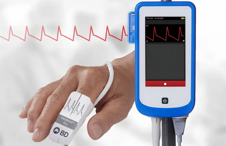
To observe real-time neurological changes, the team equipped the animals with custom, lightweight, head-mounted devices capable of simultaneously capturing electroencephalography (EEG) and electromyography (EMG) data.
- The EEG Function: By recording electrical voltage fluctuations across brain networks, the EEG provided an exact "electrical fingerprint" of neural oscillations.
- The EMG Function: By tracking muscle tone, the EMG allowed researchers to differentiate between active waking states, light non-rapid eye movement (NREM) sleep, deeply restorative NREM sleep, and rapid eye movement (REM) dreaming sleep.
Furthermore, to visualize the spatial distribution of the brain’s immune cells in relation to the plaques, the team deployed light sheet microscopy. This advanced imaging technique renders opaque brain tissue optically transparent, enabling a laser light plane to construct a high-resolution, three-dimensional digital map of the entire organ.
Identifying the "Ceiling Effect" of Early Plaques
As the investigation progressed, the team uncovered an unexpected neurobiological phenomenon that challenged conventional assumptions about disease progression. Dr. Constantino and Dr. Macauley had initially hypothesized that as plaque burden grew heavier over time, sleep disruptions would progressively worsen in a linear, compounding decline.
Instead, the data revealed a striking "ceiling effect." The disruptions in sleep architecture and cortical EEG activity observed at six months—when plaques first began to emerge—did not worsen by 18 months, despite the animals accumulating more than double the total volume of amyloid plaques.
This unexpected finding suggested that the initial wave of microglial activation triggered by early-stage plaque deposition is sufficient to establish a permanent sleep deficit. Once the inflammatory cascade is initiated, accumulating additional plaques does not produce a proportional increase in sleep disruption, indicating that the immune response operates as an independent driver of pathology.
Pharmacological Depletion and Sleep Restoration
To confirm whether microglia were directly responsible for the observed sleep deficits, the researchers administered a specialized small-molecule drug known as Pexidartinib (PLX3397). Originally developed in oncological research, PLX3397 blocks the colony-stimulating factor 1 receptor (CSF1R) signaling pathway, which microglia depend upon for survival.
After feeding the mice the drug-infused diet for 14 consecutive days, the researchers successfully depleted approximately 87% of the brain’s resident immune cells. The results were immediate and profound. Following microglial depletion, the Alzheimer’s model mice regained more than two hours of total sleep per day.
Critically, this dramatic recovery in sleep duration occurred without any change in the total volume of amyloid plaques in the brain. This proved definitively that the sleep loss was not a direct mechanical consequence of the plaques themselves, but rather the result of the inflammatory signaling storms produced by the immune cells reacting to them.
Supporting Context & Metrics
The implications of the UK study extend far beyond basic neurobiology, touching upon critical questions regarding bioenergetics, restorative sleep cycles, and accessible diagnostic biomarkers.
Dissecting Periodic vs. Aperiodic Brain Activity
To analyze the EEG data with maximum precision, the team applied an advanced mathematical algorithm known as Fitting Oscillations and One Over Frequency (FOOOF). This computational approach decomposes complex electrical brain signals into two distinct components:
- Periodic Activity: Traditional, rhythmic brain waves (such as Delta, Theta, Alpha, and Beta waves) associated with specific cognitive states and sleep stages.
- Aperiodic Activity: The background broadband electrical noise, which reflects the overall excitation-to-inhibition balance of the neural network.
Using an automotive analogy, the researchers likened this metric to evaluating whether a car’s engine continues to idle at a dangerously high, roaring speed even when the vehicle is parked. The aperiodic data confirmed that in Alzheimer’s models, the brain’s neural networks remained locked in an abnormally high state of metabolic arousal—as if the microglia were keeping the engine perpetually revved.
The Selective Targeting of NREM Sleep
A vital contribution of the study was its ability to differentiate the sleep architecture changes caused by normal aging from those driven specifically by Alzheimer’s pathology:
- Normal Aging: Primarily degraded REM (Rapid Eye Movement) sleep, the stage heavily implicated in emotional processing and memory consolidation.
- Alzheimer’s Pathology: Selectively devastated NREM (Non-Rapid Eye Movement) sleep, the deeply restorative phase characterized by slow-wave brain activity.
+-------------------------------------------------------------------------+
                    THE ALZHEIMER'S FEED-FORWARD LOOP
+-------------------------------------------------------------------------+
                                      |
                                      v
                      [ Amyloid-Beta Plaques Form ]
                                      |
                                      v
                     [ Microglia Activate & Inflame ]
                                      |
                                      v
                 [ Loss of Deep NREM Restorative Sleep ]
                                      |
                                      v
              [ Impaired Glymphatic Clearance of Toxins ]
                                      |
                                      v
              [ Accumulation of Metabolic Waste & Damage ] --+
                                                             |
+------------------------------------------------------------+
NREM sleep acts as the brain’s primary maintenance and waste-disposal window. During this phase, the glymphatic system flushes out metabolic toxins, clearing away daily cellular waste. When Alzheimer’s pathology robs patients of NREM sleep, the brain loses its primary cleaning cycle, creating a devastating feed-forward loop: poor sleep impairs waste clearance, which drives further neuroinflammation, leading to even worse sleep disruptions.
Portable EEG as an Early Diagnostic Biomarker
Recognizing that specialized neuroimaging and lumbar punctures are expensive, invasive, and inaccessible to many rural populations, the UK team envisions a future where portable, low-cost EEG headbands can be deployed in community clinics. By identifying specific electrical fingerprints that separate normal aging from early Alzheimer’s changes, clinicians could eventually screen at-risk individuals in their home environments long before clinical cognitive decline or severe memory loss becomes apparent.
Official Statements
Reflecting on the collaborative culture and rigorous intellectual environment that enabled this breakthrough, lead investigator Dr. Shannon L. Macauley emphasized the importance of curiosity-driven science and calculated risk-taking:
"Basically, we showed that it is not the plaques themselves, or solely dysfunctional neurons, that cause sleep loss but actually microglia. Microglia are immune cells that, when they respond to plaques, kick off this elaborate cascade of inflammation, as if the microglia are partying all night, and keeping the brain awake."
— Dr. Shannon L. Macauley, Associate Professor of Physiology, UK College of Medicine
Dr. Macauley also highlighted the critical role of restorative sleep in maintaining neurological health:
"That restorative sleep is super important for physical repair, learning and memory, and washing out the toxins of the day. When Alzheimer’s patients lose this stage, they lose their brain’s primary cleaning cycle, creating a feed-forward loop that may drive further damage."
First author Dr. Nicholas J. Constantino reflected on the unexpected nature of the data and the mentorship that guided the project through uncharted scientific territory:
"I expected that as plaque burden became more severe, sleep disruption would also worsen. The disruptions in sleep and cortical EEG activity that occur at six months, when plaques first emerge, did not worsen by 18 months, despite more than double the amount of plaque burden… Dr. Macauley has also taught me to embrace uncertainty and failure as part of the scientific process. Some of the most interesting studies I have been a part of emerged because our original hypothesis was wrong."
Future Outlook
Armed with these transformative insights, Dr. Macauley’s laboratory at the Sanders-Brown Center on Aging is already charting the next phase of translational research.
Rather than completely eliminating microglia—an approach that, while scientifically illuminating via Pexidartinib, could compromise normal immune surveillance in human patients—the team is pivoting toward precision immunomodulation. Current investigations are exploring whether FDA-approved medications already in clinical use, such as the diabetes drug Metformin and the antiseizure medication Stiripentol, can be repurposed to safely recalibrate microglial metabolism and quiet their inflammatory overactivity.
By successfully subduing these hyperactive immune cells without eradicating them, researchers hope to restore healthy NREM sleep architecture, interrupt the toxic feed-forward loop of neurodegeneration, and significantly improve the cognitive vitality and overall quality of life for millions of individuals living with or at risk for Alzheimer’s disease.
Funding Acknowledgments
This research was supported by the National Institute on Aging of the National Institutes of Health (Awards R01AG068330, R01AG093847, and P30AG072946), the National Institute of General Medical Sciences (Awards P30GM127211 and P20GM148326), a $287,236 award from the Cure Alzheimer’s Fund, and a $250,000 grant from The CART Fund (Coins for Alzheimer’s Research Trust).