





Executive Overview
Eosinophilic esophagitis (EoE)—a chronic, immune-mediated inflammatory condition of the esophagus driven by allergens and type 2 immune responses—has transitioned from a rare clinical curiosity into a major public health concern. Over recent decades, its incidence and prevalence have surged dramatically, with approximately 40,000 new cases diagnosed annually in the United States alone, predominantly concentrated across North America and Europe. Clinically, the burden of the disease is stark: adults routinely present with debilitating dysphagia, chest pain, and recurrent heartburn, while pediatric patients face severe feeding difficulties, vomiting, failure to thrive, and weight loss.
Left untreated or inadequately managed, chronic esophageal inflammation incites progressive tissue remodeling, leading to strictures, high-risk food impactions, and dangerous esophageal perforations. Beyond the profound physical morbidity, EoE exacts a heavy economic toll. Nationwide studies reveal that the chronic medical care of EoE patients costs more than double that of matched general populations, driven largely by frequent endoscopies, specialist consultations, and rigorous dietary or pharmaceutical interventions.
Compounding these clinical and economic challenges are systemic delays in diagnosis. On average, patients endure a diagnostic odyssey spanning more than two years from symptom onset to confirmation, suffering prolonged morbidity in the interim. Once diagnosed, therapeutic responses remain highly variable, and long-term disease monitoring relies heavily on repetitive, invasive endoscopies.
Enter artificial intelligence (AI). Over the past decade, the rapid evolution of machine learning (ML), deep learning, and natural language processing (NLP) has opened transformative frontiers in medicine. In the realm of gastroenterology—and EoE specifically—AI technologies are emerging as critical catalysts capable of addressing diagnostic delays, refining histopathological analysis, automating endoscopic scoring, predicting therapeutic responses, and revolutionizing precision medicine. This comprehensive report explores the technological evolution of AI in medicine, evaluates its current clinical applications in EoE, addresses critical limitations and ethical hurdles, and charts the regulatory roadmap for its future integration into standard clinical workflows.
Detailed Chronology: The Evolution of AI and Its Convergence with Gastroenterology
From Binary Logic to Adaptive Algorithms
The intellectual lineage of artificial intelligence traces back to the 1950s, when pioneering computer scientists engineered early systems capable of processing binary outputs and executing "if-then" statements to simulate human critical thinking. However, these early architectures were rigidly deterministic, lacking the capacity to adapt or learn from unstructured real-world variables.
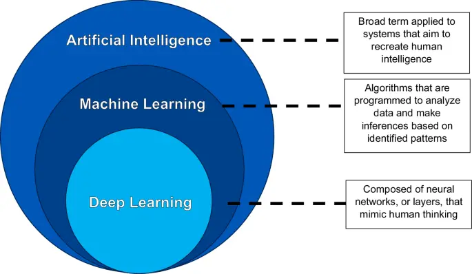
Decades of algorithmic evolution eventually gave rise to classical machine learning, wherein data is explicitly labeled and structured by human engineers, allowing models to extract patterns and formulate discrete inferences based on predefined categories. For instance, a classical ML model could be trained on distinctly labeled histologic images—such as normal squamous tissue versus glandular tissue—to identify unique features corresponding to each pre-programmed subset.
The true inflection point for modern medical informatics, however, was the advent of deep learning, a sophisticated subset of machine learning. Unlike classical ML, deep learning models require drastically less human intervention, relying instead on artificial neural networks composed of multiple hierarchical layers to independently discover and categorize data features. For example, in analyzing a tissue slide, an initial neural network layer might evaluate color gradients, a subsequent layer maps structural contours, and deeper layers synthesize these elements to independently generate outputs without explicit pre-labeling.
Within this computational hierarchy, models branch into specialized modalities:
- Computer Vision: Designed to analyze complex spatial imagery, foundational for endoscopic and histopathological assessment.
- Natural Language Processing (NLP): Engineered to parse, comprehend, and generate human language, unlocking vast electronic health record (EHR) datasets and patient education tools.
- Multimodality Models: Advanced frameworks that concurrently ingest and synthesize imaging, genetic, proteomic, and clinical data to execute complex medical tasks.
The Historical Trajectory of Medical AI
The integration of computing into clinical medicine is not entirely novel. During the 1970s and 1980s, early "diagnostic consultant" algorithms—such as INTERNIST-I and MYCIN—were developed to assist physicians in formulating differential diagnoses and tailored treatment regimens based on patient history, physical examinations, and laboratory inputs. Despite their sophisticated knowledge bases curated by medical experts, these early systems failed to achieve widespread clinical adoption. They were hindered by cumbersome interfaces, a complete lack of internet connectivity, and the absence of high-powered computational hardware.
A major paradigm shift occurred in the 2000s with the development of IBM’s Watson, which leveraged advanced natural language processing to synthesize disparate data sources at unprecedented speeds. Soon thereafter, medical-grade AI platforms emerged, capable of interpreting high-resolution radiological and endoscopic imaging, paving the way for targeted applications in chronic inflammatory disorders like EoE.
Supporting Context & Metrics: Unlocking EoE Through Data-Driven Innovation
Diagnostic Approaches and Proteomic Integration
The definitive diagnosis of EoE has traditionally relied on histopathologic criteria—specifically, the identification of greater than 15 eosinophils per high-powered field (HPF) on esophageal mucosal biopsies. Yet, clinical ambiguity frequently arises, particularly when differentiating EoE from overlapping etiologies such as gastroesophageal reflux disease (GERD), which can share both endoscopic and clinical features.
To resolve these diagnostic gray areas, cutting-edge researchers are pairing clinical data with advanced computational frameworks. In a landmark study, investigators deployed a deep learning model incorporating proteomics data derived from liquid chromatography-mass spectrometry across patient tissue slides, combined with pathologist-interpreted histologic parameters. This hybrid AI-assisted morphoproteomic approach achieved a staggering 100% diagnostic accuracy, with sensitivity and specificity exceeding 95% for EoE identification. Such innovations highlight the potential of AI to transcend human perceptual limits, establishing objective molecular signatures for complex esophageal diseases.
Case Identification and Risk Prediction
Because EoE symptoms are frequently non-specific (such as intermittent dysphagia or atypical chest pain), patients often endure long intervals before undergoing diagnostic endoscopy. Identifying individuals at risk prior to severe fibrotic complications—such as food impactions requiring emergency endoscopic intervention—is a major clinical priority.
To bridge this gap, clinical researchers developed and validated machine learning tools leveraging cohorts of adult patients reporting dysphagia. A predictive model incorporating simple clinical and demographic variables achieved an impressive Area Under the Curve (AUC) of 0.90 for predicting histologically confirmed EoE. When extended to include endoscopic inputs, the model’s predictive accuracy rose to an AUC of 0.94. To maximize clinical utility, the developers made these machine learning calculators publicly accessible online, empowering frontline clinicians to perform point-of-care risk stratification.
Pathophysiological Insights via Biomechanical Modeling
Chronic inflammation in EoE drives progressive tissue remodeling and esophageal wall stiffening. Functional luminal imaging panometry (FLIP) evaluates these mechanical properties, measuring the distensibility plateau of the esophageal wall—a metric that is markedly compromised in active EoE patients.
Using FLIP topography data from hundreds of participants, researchers constructed a virtual disease landscape utilizing mechanics-informed machine learning. This deep learning model successfully isolated distinct biomechanical profiles capable of differentiating EoE from other motility and inflammatory disorders, such as achalasia and GERD. By quantifying parameters like esophageal wall stiffness, contraction synchrony, and muscle relaxation patterns, the model offers a window into the dynamic pathophysiology of the esophagus, laying the groundwork for objective disease staging and prognosis.
Official Statements and Empirical Findings: Endoscopic, Histopathologic, and Biomarker Breakthroughs
Endoscopic Enhancements and the EREFS Framework
During routine endoscopies, busy clinicians frequently miss the subtle mucosal lesions characteristic of early EoE. To mitigate interobserver variability and operator subjectivity, computer-aided diagnostic (CADx) systems driven by convolutional neural networks (CNNs) have been deployed.
In robust validation studies, CNN models analyzing static endoscopic images have achieved exceptional performance metrics. For example, Okimoto et al. reported a CADx model with an accuracy of 94.7% and an AUC of 0.995 in distinguishing EoE patients from normal controls. Similarly, Guimaraes and colleagues developed a deep-learning architecture that achieved 91.5% accuracy and an AUC of 0.97 on external validation cohorts, successfully isolating hallmark diagnostic features such as mucosal rings, white exudates, and longitudinal furrows.
Furthermore, standardized grading systems like the EREFS score (evaluating Edema, Rings, Exudates, Furrows, and Strictures) are officially endorsed by professional clinical guidelines. Rommele et al. engineered a deep-learning model evaluated against both overall EoE diagnoses and expert EREFS scoring. The algorithm matched or exceeded the diagnostic accuracy and sensitivity of beginner endoscopists and senior fellows, achieving a 93.0% accuracy for EoE detection and 95.0% congruency with expert EREFS scoring.
Revolutionizing Histopathology and Spatial Biology
Microscopic evaluation remains the gold standard for EoE diagnosis and monitoring, yet manual eosinophil counting is notoriously labor-intensive and subject to interobserver fatigue. While the Eosinophilic Esophagitis Histologic Scoring System (EoEHSS) offers superior prognostic granularity compared to simple peak eosinophil counts (PEC), its widespread clinical adoption has been sluggish due to its cumbersome nature.
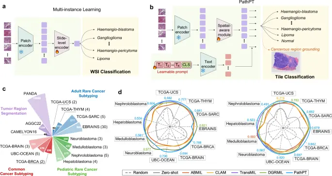
Artificial intelligence offers a definitive solution through automated digital pathology. Archila et al. engineered an AI digital pathology model trained on esophageal biopsies spanning the full spectrum of inflammatory severity. The platform successfully segmented and quantified histologic features—including epithelial layers, intercellular edema, granules, and specific cell counts—performing on par with experienced gastrointestinal pathologists. Across large validation cohorts, the model retained an AUC of 0.89 for evaluating PEC, alongside high reproducibility.
Complementing this, advanced machine learning platforms have begun mapping spatial biology within the esophageal microenvironment. Studies evaluating mast cell dynamics have utilized AI to uncover significant increases in epithelial mast cell infiltration and degranulation in active EoE compared to healthy controls, shedding light on underlying pathogenic mechanisms driven by mediators like interleukin-13 (IL-13).
Biomarkers and the Promise of Precision Medicine
Despite extensive research, a universally accepted non-invasive biomarker for diagnosing EoE or monitoring therapeutic response remains elusive. Microarray analyses and machine learning integration have identified novel candidate genes within the MUC and SPRR families linked to EoE pathogenesis. Furthermore, machine learning classification of microRNA (mRNA) expression profiles from pediatric cohorts has demonstrated exceptional diagnostic accuracy (AUC: 98.5), successfully identifying cryptic cases and tracking therapeutic responses following topical corticosteroid therapy.
Future Outlook: Clinical Hurdles, Ethical Frameworks, and Regulatory Pathways
Overcoming Current Limitations
Despite the boundless optimism surrounding medical AI, clinical translation must proceed with rigorous caution. Several systemic limitations threaten the safe integration of these technologies:
- Sampling Bias and Generalizability: Many current EoE models are trained on relatively small datasets comprising fewer than 200 patients. Models trained on homogenous populations risk severe overfitting and may fail to generalize across diverse racial, ethnic, and socioeconomic demographics.
- The "Black Box" Dilemma: Many deep-learning algorithms generate high-stakes clinical predictions without transparently communicating the underlying reasoning. In clinical practice, where "clinical gestalt" and human intuition reign supreme, the demand for explainable artificial intelligence (XAI) is paramount.
- Ethical Accountability and Liability: When an AI algorithm misclassifies an esophageal biopsy or fails to flag a pre-cancerous lesion, questions of legal liability immediately surface. Determining whether fault lies with the software developer, the attending physician, or the healthcare institution remains an unresolved legal frontier.
- Generative AI Inaccuracies: Evaluations of general-purpose large language models (such as ChatGPT) in answering patient-centric EoE questions have revealed persistent scientific inaccuracies. Notably, un-tuned chatbots have generated alarming misinformation—such as falsely claiming a direct link between EoE, Barrett’s esophagus, and esophageal adenocarcinoma—contrary to large-scale epidemiological data demonstrating that EoE does not elevate esophageal cancer risk.
Regulatory Oversight and AI-SaMD
As artificial intelligence embeds deeper into clinical workflows, appropriate regulatory frameworks are essential to safeguard patient health. In the medical technology sector, AI applications are officially classified as Artificial Intelligence-based Software as a Medical Device (AI-SaMD). This category encompasses diagnostic imaging algorithms, clinical decision support software, automated histopathology tools, and remote monitoring platforms.
Unlike traditional static medical software, AI-SaMD possesses the unique capacity for continuous learning and algorithmic evolution over time. In the United States, the Food and Drug Administration (FDA) regulates AI-SaMD through a risk-based classification framework:
- Class I (Low Risk): Subject to general controls.
- Class II (Moderate Risk): Requires special controls and premarket notification (510(k) clearance).
- Class III (High Risk): Requires rigorous premarket approval (PMA) due to significant potential impact on patient morbidity and mortality.
Regulatory bodies face the dual challenge of enforcing stringent safety and efficacy standards while maintaining flexible review pathways that do not stifle rapid technological innovation.
Conclusion: Moving Toward Precision Care in EoE
The integration of artificial intelligence into the management of eosinophilic esophagitis marks a profound turning point in modern gastroenterology. By bridging diagnostic delays, automating tedious histopathological and endoscopic scoring, and decoding complex biomechanical and molecular datasets, AI holds the key to transitioning EoE care from a reactive, trial-and-error paradigm into a proactive, personalized precision medicine framework. While significant hurdles regarding data diversity, algorithm transparency, and liability must be systematically addressed, the continued maturation of AI promises a future of earlier diagnoses, streamlined treatments, and vastly improved quality of life for patients worldwide.