







Executive Overview
In a stunning revelation that challenges foundational assumptions about cellular biology and immunology, a team of researchers at Stanford University has identified a previously unknown type of immune cell that destroys neighboring threats by literally blowing itself apart. The process—occurring with such astonishing velocity and totality that the cell vanishes completely within minutes—leaves virtually no trace behind, fundamentally expanding the known spectrum of programmed and reactive cell death.
Published in the premier scientific journal Cell, the discovery centers on planarian flatworms (Schmidtea mediterranea), small aquatic organisms globally celebrated for their virtually limitless capacity for regeneration. When investigators investigated how these creatures distinguish their own biological material from foreign tissue, they stumbled upon an unprecedented defensive mechanism. Named "ruptoblasts" by the research team, these glandular cells respond to a specific hormonal trigger by transforming into microscopic biological bombs.
The implications of this discovery stretch far beyond the biology of flatworms. By demonstrating an evolutionary defense strategy entirely distinct from the hematopoietic immune cells (such as T cells and neutrophils) found in vertebrates, this study opens new frontiers for synthetic biology, oncology, and pharmacology. Although humans and other mammals lack this explosive capability—likely sacrificed over evolutionary history due to the catastrophic collateral damage it would inflict on tissues lacking regenerative prowess—understanding the mechanics of "ruptosis" could inspire revolutionary targeted therapies for recalcitrant bacterial infections and treatment-resistant tumors.
Detailed Chronology of the Discovery
Investigating the "Frankenstein" Worms
The journey toward this groundbreaking discovery began with a fundamental question in invertebrate biology: Are planarian flatworms capable of self-recognition, and how do they mount an immune response against foreign intrusions?
Dr. Chew Chai, a postdoctoral researcher in the laboratory of Stanford bioengineering professor Bo Wang, set out to solve this long-standing mystery. To observe how flatworms handle foreign tissue, Chai engineered a series of chimeric specimens. Using micro-surgical precision, she sliced worms lengthwise and fused each half with the corresponding half of an entirely different, unrelated flatworm.
While planarians are world-renowned for their ability to regenerate missing heads, tails, and internal organs from microscopic fragments, these fused "Frankenstein" worms exhibited a violent aversion to one another. Rather than integrating peacefully, the combined organisms rejected the foreign tissue entirely. The visual and physiological parallels to mammalian organ transplant rejection were immediately striking, yet the underlying cellular choreography bore no resemblance to anything documented in human or murine models.
"It’s this huge inflammatory response," Chai explained, describing the catastrophic tissue breakdown. "Like there’s a fire and an alarm goes off, and the cells just blow up."
The Hormonal Catalyst: Activin
To pinpoint the molecular driver of this violent rejection, Chai examined the hormonal shifts occurring within the fused flatworms. Previous scholarship into planarian regeneration had highlighted the vital role played by activin, a member of the transforming growth factor-beta (TGF-$beta$) superfamily. In healthy planarians, activin maintains a delicate equilibrium: elevated levels can impede the organism’s capacity to regenerate tissues, while depressed levels sabotage its reproductive viability.
As Chai monitored the tissue-fusing experiments, she detected a sharp, localized surge in activin right as the organisms began rejecting the foreign cells. This surge was immediately followed by a cascade of chronic inflammation that, while not immediately fatal to the host organism, proved lethal within several days if the tissue conflict remained unresolved.
To confirm whether activin served as the direct catalyst for this inflammatory chaos, Chai administered controlled injections of the hormone directly into healthy, non-fused flatworms. The results were instantaneous and definitive: the introduction of activin alone was sufficient to trigger the identical, catastrophic inflammatory cascade observed in the chimeric specimens.
Capturing "Ruptosis": Cell Death in Minutes
Determined to witness the cellular mechanics driving this reaction, Chai deployed advanced live-cell microscopy alongside flow cytometry—a high-throughput laser-based technique capable of sorting and analyzing heterogeneous cell populations. By tagging cellular components with specialized fluorescent dyes, she isolated the specific subpopulation of cells that reacted to the activin surge.
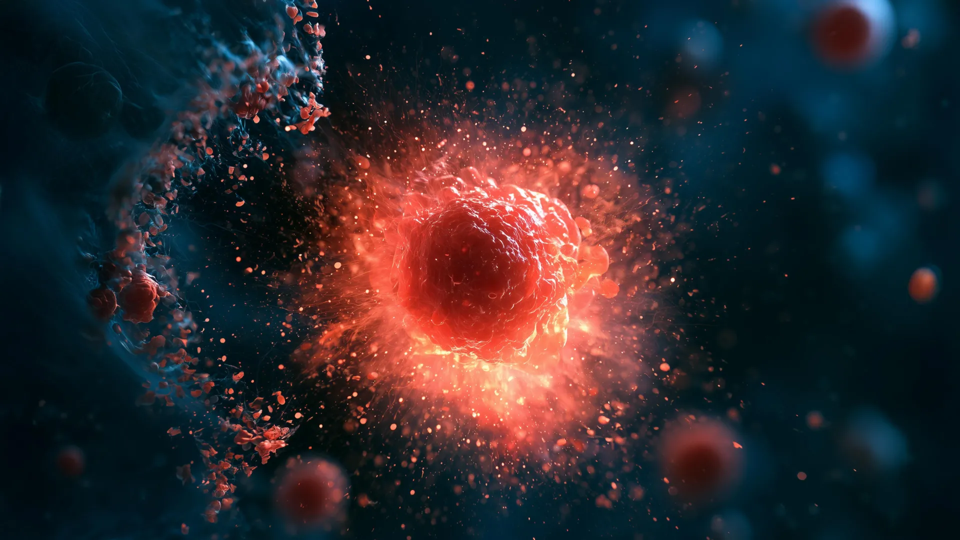
What the microscope revealed defied conventional paradigms of cell death. Rather than undergoing apoptosis (programmed cell death characterized by orderly disassembly and phagocytosis) or necrosis (passive, messy cell lysis due to acute injury), a small group of glandular cells suddenly underwent rapid, violent hyper-distension. Within a window of five minutes, these cells breached their own membranes, forcefully jettisoning a payload of toxic internal substances that instantly eradicated adjacent cells in the immediate microenvironment.
Recognizing that this phenomenon was entirely distinct from existing classifications of cellular demise, Chai and Wang coined a new term for the process: ruptosis.
Supporting Context & Metrics: How Ruptosis Redefines Cell Death
To understand the radical nature of ruptosis, it is helpful to place it within the context of established biological paradigms. Cell death in biological systems has traditionally been categorized into neatly defined pathways, each operating on specific timescales and serving distinct physiological roles.
| Cell Death Mechanism | Primary Triggers | Timescale | Nature of Destruction | Collateral Impact |
|---|---|---|---|---|
| Apoptosis | Developmental cues, internal DNA damage | Hours | Controlled disassembly, apoptotic bodies | Minimal; cleared quietly by phagocytes |
| Necrosis | Severe physical trauma, ischemia | Minutes to hours | Passive membrane rupture, swelling | High; triggers widespread inflammation |
| NETosis (Neutrophils) | Pathogen encounter | Hours | Extrusion of DNA traps (NETs) | Moderate; localized antimicrobial defense |
| Ruptosis (Ruptoblasts) | Activin surge, foreign tissue detection | Seconds to minutes | Explosive, complete, active self-destruction | Highly localized; neutralizes targets within micron-range |
Mechanics of the Explosion
The velocity of ruptosis separates it entirely from other known explosive cellular behaviors. While certain mammalian cells and bacterial pathogens can undergo explosive lysis—such as inflammasome-driven pyroptosis or neutrophil extracellular trap (NET) formation—these processes are comparatively sluggish.
"Some mammalian cells and bacteria may also do an explosive sort of cell death, but the timescale is really long," Chai noted. "They are exploding, but it’s more like pores that slowly leak things out over the course of several hours. Ruptosis happens within seconds to minutes."
This compression of timeline transforms the ruptoblast from a passive secretory vessel into an aggressive, ballistic weapon. Instead of leaking chemicals gradually, the cell unloads its entire biochemical arsenal instantaneously. Mechanistically, the researchers discovered that this explosive release is powered by a massive, sudden surge of calcium ions ($textCa^2+$) mobilized directly from the endoplasmic reticulum housed within the ruptoblast itself.
Cross-Species Target Validation
To test the efficacy and lethality of these specialized cells, the Stanford team exposed isolated ruptoblasts to a diverse array of biological targets in vitro. The results demonstrated broad-spectrum cytotoxicity:
- Bacterial Pathogens: Tested against Escherichia coli (E. coli), ruptoblasts successfully obliterated bacterial colonies within their blast radius.
- Mammalian Cells: Human kidney cells and murine blood cells introduced to the explosive discharge suffered immediate and total cellular destruction.
Crucially, the destructive capacity of ruptosis is strictly localized. The blast radius remains confined to the immediate micron-scale vicinity of the exploding ruptoblast. The reaction does not propagate via a chain reaction, nor does it leave behind persistent chemical toxicity once the immediate payloads are dissipated. This high-precision, low-residual profile has electrified bioengineers seeking novel tools for localized tissue sterilization.
Official Statements and Research Perspectives
The discovery, detailed extensively in Cell, underscores the immense value of venturing outside traditional, mammalian-centric laboratory models. While mice, fruit flies (Drosophila melanogaster), and zebrafish dominate modern biomedical research, the natural world harbors an astonishing diversity of immune architectures shaped by hundreds of millions of years of distinct evolutionary pressures.
"We never expected that a cell could just explode like a bomb and kill the cells surrounding it."
— Dr. Bo Wang, Senior Author and Associate Professor of Bioengineering, Stanford University
Wang emphasizes that the invisibility of ruptoblasts in higher vertebrates is likely a matter of evolutionary risk management. Vertebrates possess complex, highly integrated organ systems where uncontrolled, explosive cellular blasts would cause catastrophic, irreparable damage to surrounding healthy tissue. Flatworms, conversely, operate under an entirely different biological economy. Backed by an inexhaustible pool of adult stem cells (known as neoblasts), planarians can effortlessly replace massive swaths of tissue lost to internal warfare.
"It demonstrates there’s lots of different immune mechanisms out there. There’s all these animals that live in an environment where there’s lots of bacteria, lots of viruses, and we know so little about their immune mechanisms."
— Dr. Chew Chai, Postdoctoral Researcher and Lead Author, Stanford University
Co-investigators point out that ruptoblasts represent an entirely novel lineage of immune actors. Unlike traditional immune sentinels such as T cells, B cells, and neutrophils—which originate from hematopoietic stem cells within the bone marrow—ruptoblasts are classified as glandular cells. They co-opt and hyper-intensify normal intracellular secretion machinery, transforming routine glandular pathways into hair-trigger explosive devices upon detecting activin signaling.
Future Outlook: Translating Ancient Biology into Modern Medicine
While flatworms and humans are separated by vast phylogenetic distances, the discovery of ruptosis offers tantalizing clues for translational medicine. As researchers look toward the horizon, several compelling pathways for clinical and technological application emerge:
- Targeted Oncology and Tumor Ablation: Solid tumors often create immunosuppressive microenvironments that evade standard immunotherapies. By understanding how ruptoblasts deliver a potent, tightly contained destructive payload without triggering systemic toxicity, bioengineers hope to design synthetic, activin-triggered micro-actuators or engineered cellular proxies that can home in on cancerous masses and self-destruct within the core of a tumor.
- Combating Antimicrobial Resistance: With antibiotic resistance representing one of the gravest public health threats of the 21st century, exploring non-traditional antimicrobial mechanisms is paramount. The ability of ruptoblast contents to eradicate resilient pathogens like E. coli points toward the development of novel, bio-inspired bactericidal agents capable of breaching tough bacterial cell walls through rapid chemical shock.
- Broadening the Scope of Evolutionary Immunology: The research team’s phylogenetic screening revealed that cells functionally analogous to ruptoblasts appear restricted to basal bilaterians—ancient evolutionary branches that split from the human lineage hundreds of millions of years ago. This suggests that mammalian lineages discarded this mechanism as their regenerative architectures evolved. Re-examining "non-model" organisms may unveil a treasure trove of forgotten biological innovations waiting to be harnessed.
As the Stanford team continues to dissect the precise molecular cascades governing calcium flux and membrane destabilization during ruptosis, the scientific community is reminded that nature’s ingenuity far outstrips human imagination. By listening to the quiet, resilient biology of flatworms, modern medicine may soon borrow a page from an ancient defense strategy, turning cellular self-destruction into a powerful tool for healing.
Acknowledgements & Funding
This foundational research was conducted by an interdisciplinary team at Stanford University and Ben-Gurion University of the Negev. Key Stanford co-authors include postdoctoral scholar Souradeep Sarkar; former Undergraduate Visiting Research Program scholar Lihan Zhong; Dania Nanes Sarfati, PhD ’24; Christine Jacobs-Wagner, Dennis Cunningham Professor of Biology and Microbiology & Immunology; and Hawa Racine Thiam, Assistant Professor of Bioengineering and Microbiology & Immunology. Co-senior author Benyamin Rosental represented Ben-Gurion University.
Institutional support was provided by Stanford Bio-X, the Wu Tsai Neurosciences Institute, the Maternal & Child Health Research Institute (MCHRI), and Sarafan ChEM-H. Financial backing for the project was granted by the National Science Foundation (Graduate Research Fellowship), Stanford University (Graduate and DARE fellowships), the Human Frontier Science Program, the National Institutes of Health (NIH), and the European Research Council.