











Executive Overview
In the intricate landscape of hematology, pancytopenia—a dangerous reduction in red blood cells, white blood cells, and platelets—serves as an urgent diagnostic alarm. It typically commands an immediate, exhaustive clinical workup to unmask life-threatening underlying conditions such as bone marrow failure syndromes, hematologic malignancies, or profound nutritional deficiencies.
However, a landmark case report published in Frontiers in Hematology (April 2026) illuminates a sophisticated and perilous diagnostic trap. The report details the clinical journey of a 67-year-old Latina patient who endured a grueling two-year medical odyssey. Initially misdiagnosed with myelodysplastic syndrome (MDS) coupled with an immune-mediated hemolytic process, she experienced recurrent cycles of pancytopenia that repeatedly and deceptively responded to aggressive immunotherapy, including corticosteroids and the anti-CD20 monoclonal antibody rituximab.
This unusual temporary responsiveness to immunotherapy—combined with normal standard laboratory test results for vitamin $textB12$—masked the true culprit: pernicious anemia. High titers of anti-intrinsic factor antibodies not only drove her autoimmune pathology but also interfered with standard luminescence binding assays, generating falsely normal vitamin $textB12$ readings. This case exposes vulnerable blind spots in contemporary hematological diagnostics. It underscores how autoimmune features can mimic aggressive bone marrow disorders, leading to diagnostic misclassification, profound psychological trauma for the patient, and protracted exposure to unnecessary, potent pharmacological agents.
Detailed Chronology: A Two-Year Diagnostic Labyrinth
Presentation and Initial Workup
The case centers on a 67-year-old woman with a medical history notable for type II diabetes mellitus, essential hypertension, and thyroid nodules. Prompted by acute pancytopenia discovered during an emergency department visit, she presented to a hematology clinic reporting a gradual, debilitating onset of severe fatigue, exertional dyspnea, easy bruising, and chest wall discomfort. She exhibited no classic "B" symptoms (fever, night sweats, unexplained weight loss) or acute neurological deficits.
Initial laboratory evaluations revealed profound cytopenias and striking markers of active hemolysis:
- White Blood Cell Count (WBC): $2.6 times 10^9/textL$ (Absolute Neutrophil Count: $0.9text K/mutextL$)
- Hemoglobin (Hb): $76text g/L$ (Hematocrit: $20.3%$)
- Mean Corpuscular Volume (MCV): $100.9text fL$
- Platelets: $90 times 10^9/textL$
- Total Bilirubin: $2.6text mg/dL$ (Indirect/Unconjugated: $2.0text mg/dL$)
- Lactate Dehydrogenase (LDH): $5,575text U/L$ (Reference: $90text–180text U/L$)
- Haptoglobin: $<10text mg/dL$ (Reference: $30text–200text mg/dL$)
- Reticulocyte Index: $0.57$
Despite overwhelming hemolytic indicators, both standard and enhanced direct antiglobulin tests ("Super Coombs") were entirely negative. Serum folate was normal ($14.8text ng/mL$), and her total serum vitamin $textB_12$ level returned at a seemingly reassuring $410text pg/mL$ (Reference range: $181text–914text pg/mL$).
A bone marrow biopsy revealed prominent dyserythropoiesis alongside myeloid and megakaryocyte dysplasia, though blasts and ring sideroblasts were not initially increased. Comprehensive molecular and cytogenetic evaluations—including myeloid FISH panels, 63-gene NGS panels, and a 115-gene bone marrow failure panel—yielded entirely negative results, leaving clinicians without a definitive genetic signature for primary MDS.
The Immunotherapy Loop and Deceptive Responses
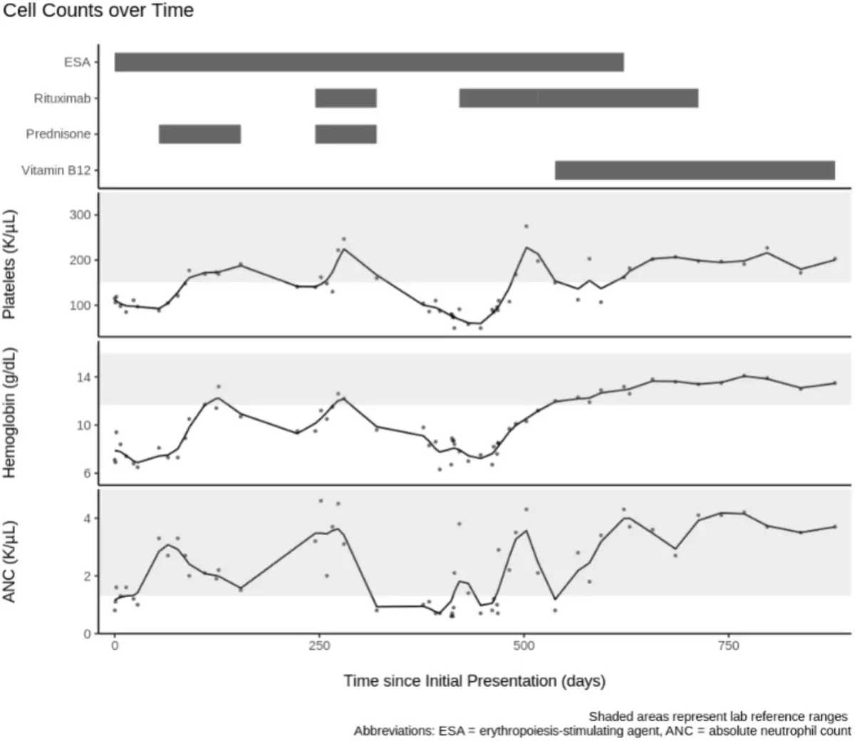
Given the clinical suspicion of MDS paired with an atypical immune-mediated hemolytic phenomenon, clinicians initiated a course of oral prednisone ($1text mg/kg$ daily) alongside exogenous erythropoietin. Remarkably, the patient achieved a complete resolution of her cytopenias within four months.
However, three months after tapering off prednisone, her pancytopenia relapsed ($textHb 95text g/L$, $textPlt 140 times 10^9/textL$) alongside biochemical signs of recurrent hemolysis. Interpreting this as refractory immune-mediated cytopenia, medical teams resumed high-dose prednisone and administered four weekly infusions of rituximab ($375text mg/m^2$). Once again, her blood counts rebounded significantly.
Escalation, Relapse, and Morphological Shift
Four months later, the patient suffered a third recurrence, accompanied by an unintentional $3text kg$ weight loss. Comprehensive imaging via CT scans of the chest, abdomen, and pelvis showed no lymphadenopathy or organomegaly, and paroxysmal nocturnal hemoglobinuria flow cytometry remained negative.
A repeat bone marrow examination revealed a dramatic morphological evolution:
- Trilineage dysplasia now affected over $20%$ of elements in each lineage.
- Erythroid elements exhibited marked megaloblastic maturation with numerous binucleated forms.
- Megakaryocytes displayed small, hyposegmented structures, and granulocytes featured aberrant nuclear segmentation.
- Iron staining demonstrated that ring sideroblasts accounted for $11%$ of erythroid elements.
Despite these alarming morphological findings that strongly simulated high-risk MDS, repeat cytogenetic and NGS studies remained entirely negative.
Faced with mounting clinical deterioration, clinicians initiated a second cycle of rituximab (five weekly doses followed by eight monthly maintenance infusions), supplemented by another course of prednisone when counts plunged yet again. Each intervention temporarily revived her hematologic indices, creating a deceptive clinical illusion that an immune-driven hematologic malignancy was being managed.
Unmasking the Truth: Nutritional Salvage
Faced with non-sustained responses to aggressive immunosuppression and persistently perplexing bone marrow morphology, the clinical team re-evaluated nutritional etiologies.
When vitamin $textB_12$ was re-tested, the value had plummeted to $101text pg/mL$. More critically, serum methylmalonic acid (MMA) testing revealed a staggering elevation at $29 mutextmol/L$ (Reference: $0.00text–0.40 mutextmol/L$). Subsequent testing for anti-intrinsic factor antibodies (anti-IF Ab) returned positive, confirming the true underlying pathology: pernicious anemia.
Immunosuppressive therapy was immediately halted, and the patient was transitioned to parenteral vitamin $textB_12$ replacement. The response was swift, complete, and enduring. At her 28-month follow-up evaluation, her blood counts remained entirely normalized, and she was clinically thriving. Surveillance endoscopies revealed gastric intestinal metaplasia, which remains under close gastroenterological surveillance.
Supporting Context & Metrics
| Diagnostic Parameter | Initial Presentation | Third Relapse | Post-Parenteral $textB_12$ Therapy |
|---|---|---|---|
| Hemoglobin (Hb) | $76text g/L$ | $85text g/L$ | Normal ($>120text g/L$) |
| MCV | $100.9text fL$ | $90.6text fL$ | Normal Range |
| Platelets | $90 times 10^9/textL$ | $74 times 10^9/textL$ | Normal ($>150 times 10^9/textL$) |
| Lactate Dehydrogenase (LDH) | $5,575text U/L$ | $9,413text U/L$ | Normalized |
| Serum Vitamin $textB_12$ | $410text pg/mL$ (Spuriously Normal) | $101text pg/mL$ (Deficient) | Repleted / Maintained |
| Methylmalonic Acid (MMA) | Not Performed | $29 mutextmol/L$ (Critically High) | Normalized |
| Anti-Intrinsic Factor Antibodies | Not Performed | Positive | Positive |
The Mechanics of Assay Interference
The core driver of this two-year diagnostic delay lies in the biochemical limitations of standard laboratory assays. The vast majority of clinical laboratories measure serum vitamin $textB12$ using rapid competitive luminescence binding assays. In these tests, labeled vitamin $textB12$ tracers compete with endogenous patient $textB_12$ to bind with exogenous intrinsic factor supplied in the assay kit.
In patients with pernicious anemia, high titers of circulating anti-intrinsic factor antibodies bind to the assay’s intrinsic factor reagent. This blocks the binding of the labeled tracer, paradoxically generating spuriously normal or elevated $textB_12$ readings. While modern assays incorporate denaturation steps to neutralize these antibodies, high-titer variants frequently overwhelm these protocols, leaving clinicians falsely reassured.
Why Did Immunotherapy Work?
The patient’s clear-cut clinical responses to prednisone and rituximab represented a major confounding factor. Pernicious anemia is fundamentally an autoimmune disease driven by autoantibody-mediated destruction of gastric parietal cells and intrinsic factor. Experts hypothesize that broad immunosuppressive regimens partially suppressed the underlying autoimmune-driven pathophysiology, transiently mitigating ineffective erythropoiesis and improving marrow function—even though they failed to correct the core metabolic deficiency.
Official Statements & Expert Insights
Reflecting on the psychological and clinical impact of the case, corresponding author Dr. John G. Quigley and his colleagues at the University of Illinois Chicago emphasized the severe toll of diagnostic misclassifications:
"As occurred here, a misdiagnosis of myelodysplastic syndrome may confer substantial emotional trauma and potentially lead to unnecessary treatments with hematopoietic growth factors and/or hypomethylating agents."
Furthermore, the authors underscored the critical takeaway regarding modern diagnostic workflows for unexplained cytopenias:
"This case underscores the potential limitations of relying solely on serum vitamin $textB12$ measurements in patients with unexplained cytopenias… If there is clinical suspicion for pernicious anemia, measurement of MMA should be considered when $textB12$ levels are normal or low-normal."
The research team also highlighted that primary MDS unconfirmed by somatic gene mutations or canonical cytogenetic aberrations is exceptionally rare ($<10%$), arguing that such presentations should consistently prompt a rigorous re-evaluation for secondary and nutritional mimics.
Future Outlook & Clinical Recommendations
The lessons drawn from this case chart a clear path forward for hematology and internal medicine practice. As diagnostic platforms evolve, clinicians must remain acutely aware that normal laboratory values do not categorically rule out metabolic deficiencies, particularly in autoimmune contexts.
- Broadening Metabolic Screening: When evaluating unexplained pancytopenia—especially when accompanied by macrocytosis or hemolytic markers—clinicians should maintain a low threshold for ordering functional metabolic markers such as serum methylmalonic acid (MMA) and homocysteine, regardless of initial serum $textB_12$ concentrations.
- Challenging Refractory "Immune" Diagnoses: Patients presenting with cytopenias that display atypical or non-sustained responses to immunosuppressive therapies should undergo comprehensive re-investigation of their baseline metabolic and autoimmune profiles.
- Recognizing MDS Mimics: Bone marrow morphology alone can be notoriously deceptive. Nutritional deficiencies can faithfully replicate the dysplastic features of myelodysplastic syndromes. Integrating exhaustive genetic panels alongside meticulous metabolic evaluation prevents patients from enduring the psychological weight and physical risks of an erroneous malignant diagnosis.
By bridging the gap between immunology and metabolic medicine, healthcare providers can prevent similar diagnostic odysgeries, sparing patients from prolonged uncertainty and ensuring targeted, curative interventions are administered swiftly.