







Executive Overview
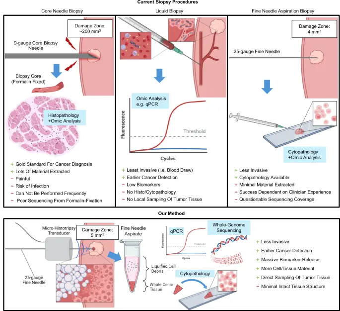
The landscape of cancer diagnostics is on the precipice of a profound transformation, driven by an innovative technique that bridges the gap between minimally invasive patient comfort and high-yield molecular pathology. Core needle biopsies (CNBs) have long stood as the gold standard for solid tumor diagnosis across the breast, prostate, liver, lung, and lymph nodes due to their high specificity and structural tissue preservation. However, the clinical utility of CNBs is systematically hindered by severe patient aversion, the necessity for larger-caliber instruments, high procedural costs, and a substantial risk profile encompassing infection, bleeding, hematoma formation, and needle tract tumor seeding.
To circumvent these barriers, researchers have developed Shock-Scattering Micro-Histotripsy-Aided Fine-Needle Aspiration Biopsy (MH+FNAB). This groundbreaking diagnostic paradigm marries non-thermal, mechanically precise ultrasound tissue ablation with fine-needle aspiration (FNA). By liquefying a mere 500 nanoliters of targeted tumor volume, MH+FNAB amplifies the release of cancer-specific DNA epimutations and mRNA biomarkers by thousands of times without compromising the overall structural integrity of the tumor. This allows for rapid, high-yield molecular characterization, cytological adequacy, and whole-genome sequencing via a procedure that produces roughly 1% of the tissue disruption associated with a standard core needle biopsy.
Detailed Chronology and Technological Evolution
The Historical Dilemma of Tissue Sampling
For decades, clinicians have walked a tightrope between diagnostic accuracy and patient morbidity. Fine-needle aspiration biopsy (FNAB) emerged historically as a highly desirable, same-day, minimally invasive option that dramatically reduces patient psychological distress and mitigates hospital burdens. Yet, its clinical prevalence plummeted with the rise of CNB due to two glaring flaws: an unacceptably high rate of sampling inadequacy—sometimes soaring past 12% in image-guided procedures—and an absolute requirement for an on-site, specialized cytopathologist to evaluate sample quality in real-time.
Concurrently, the emergence of liquid and blood biopsies offered a beacon of hope for non-invasive molecular profiling. Yet, early-stage biomarker concentrations in circulation remain stubbornly low, obscuring clinical utility. While prior attempts to leverage high-intensity focused ultrasound (FUS) and boiling histotripsy attempted to boost circulating cell-free biomarkers into the bloodstream, these techniques proved either too destructive or yielded variable, unreliable concentrations.
The Inception of Micro-Histotripsy
Histotripsy is an emerging ultrasound modality that harnesses extreme negative pressures to initiate acoustic cavitation. By generating peak negative pressures that exceed the intrinsic cavitation thresholds of biological tissues, microbubble clouds form, expand, and collapse, mechanically fractionating adjacent cells on a subcellular level without causing appreciable thermal elevation.
Traditional histotripsy systems have relied on low-frequency, massive transducer arrays designed for deep-tissue, non-invasive surgical ablation. However, the advent of micro-histotripsy—utilizing higher frequencies (>5 MHz) and compact, form-factor transducers—has shifted the paradigm toward precision diagnostics. Shock-scattering micro-histotripsy exploits non-linear propagation to generate high peak-positive-pressure shock-wavefronts. When these wavefronts phase-invert upon striking gas nucleation sites, they produce localized peak-negative pressures exceeding 30 MPa.
This results in the formation of sub-millimeter cavitation clouds (approximately 500 nL in volume) that achieve pure mechanical tissue liquefaction with a razor-sharp boundary between dead and living cells. Crucially, because the damage zone is exceptionally minute, it leaves the remainder of the tumor entirely intact, preserving the option for subsequent core needle biopsies if clinically warranted.
Supporting Context and Empirical Metrics
In preclinical evaluations utilizing murine models of breast cancer and melanoma, the MH+FNAB technique demonstrated extraordinary enhancements across multiple diagnostic dimensions:
- Epigenetic Biomarker Amplification: Targeting the hypermethylation of CpG islands in the BRCA1 promoter region—a classic epimutation indicative of both hereditary and sporadic breast cancers—MH+FNAB yielded a staggering median fold-increase of 3,174.9 for the methylated allele compared to pre-histotripsy controls. Conventional FNA performed by trained specialists failed to achieve detectable levels for these alleles.
- Cancer Subtype Profiling: In luminal breast cancer xenograft models (ER/PR+), micro-histotripsy successfully amplified estrogen receptor (ER) and progesterone receptor (PR) mRNA levels by median factors of 2,645-fold and 5,599-fold, respectively, correctly denoting hormone receptor status within minutes.
- Cytopathological Adequacy: While conventional FNA yielded insufficient cellular material in four out of five attempts, MH+FNAB consistently extracted high-cellularity lysates containing loose single cells and diagnostic clusters. Pathologist-blinded reviews confirmed classic markers of malignancy, including condensed chromatin, prominent nucleoli, and positive staining for the melanoma marker S100A4.
- Whole-Genome Sequencing (WGS): Utilizing only the supernatant fraction post-centrifugation of a 20 µL micro-histotripsy aspirate, researchers successfully extracted approximately 120 ng of high-quality genomic DNA without requiring pre-amplification. WGS successfully mapped thousands of non-synonymous exonic variants and identified multiple genes implicated in human cancers via the COSMIC database, vastly outperforming formalin-fixed paraffin-embedded (FFPE) samples prone to chemical fragmentation and cross-linking artifacts.
Official Statements and Research Insights
Principal investigators behind the breakthrough emphasize that the technology directly answers an unfulfilled clinical need for rapid, high-confidence tissue diagnostics that sidestep the trauma of major surgical or core interventions.
"Our method relies on completely mechanical shock-scattering micro-histotripsy, maintaining a steadfast focus on minimizing disturbance to the overall tumor structure," note the study’s authors. "Affecting only 500 nanoliters of volume, this technique adds negligible damage to an FNA procedure, successfully bridging the chasm between the low invasiveness of cytology and the high molecular yield of surgical pathology."
Furthermore, clinical specialists uninvolved in the immediate study have praised the approach for its potential to democratize fine-needle aspiration. By liquefying solid tissue into an easily aspirable homogenate, the technique overcomes the historical dependency on an on-site cytopathologist, transforming an operator-sensitive art into a standardized, highly repeatable clinical workflow.
Future Outlook and Clinical Translation
While the preclinical data establishing MH+FNAB are robust, researchers emphasize several critical milestones that must be cleared prior to widespread human clinical adoption:
- Panel Expansion: Transitioning from single-marker detection (such as BRCA1 hypermethylation) to comprehensive, multiplexed panels incorporating diverse gene mutations and epimutations.
- Transducer Engineering: Developing advanced transducers with steerable foci and extended focal depths (beyond the current 7 mm limit) to ensure reliable access to deeper anatomical structures, such as deep-seated hepatic or pancreatic lesions. Alternatively, needle-tip histotripsy technologies utilizing flexural modes offer promising pathways to extend treatment depths using standard 25-gauge needles.
- Safety and Metastatic Profiling: Conducting exhaustive longitudinal studies to definitively rule out theoretical risks of intravascular dissemination or metastatic seeding, building upon existing histotripsy literature that points toward favorable anti-tumor systemic immune (abscopal) responses.
- Clinical Workflow Integration: Standardizing the pairing of compact micro-histotripsy driver boards with ubiquitous clinical ultrasound imaging hardware (such as VisualSonics platforms), thereby minimizing capital expenditure hurdles for hospital adoption.
If successfully translated to human trials, shock-scattering micro-histotripsy-aided fine-needle aspiration promises to rewrite clinical guidelines. By delivering rapid, inexpensive, multi-modal diagnostics—spanning cytopathology, quantitative PCR, and whole-genome sequencing—from a virtually painless, pin-prick procedure, oncology may finally move past the false dichotomy between patient comfort and diagnostic certainty.