





Executive Overview
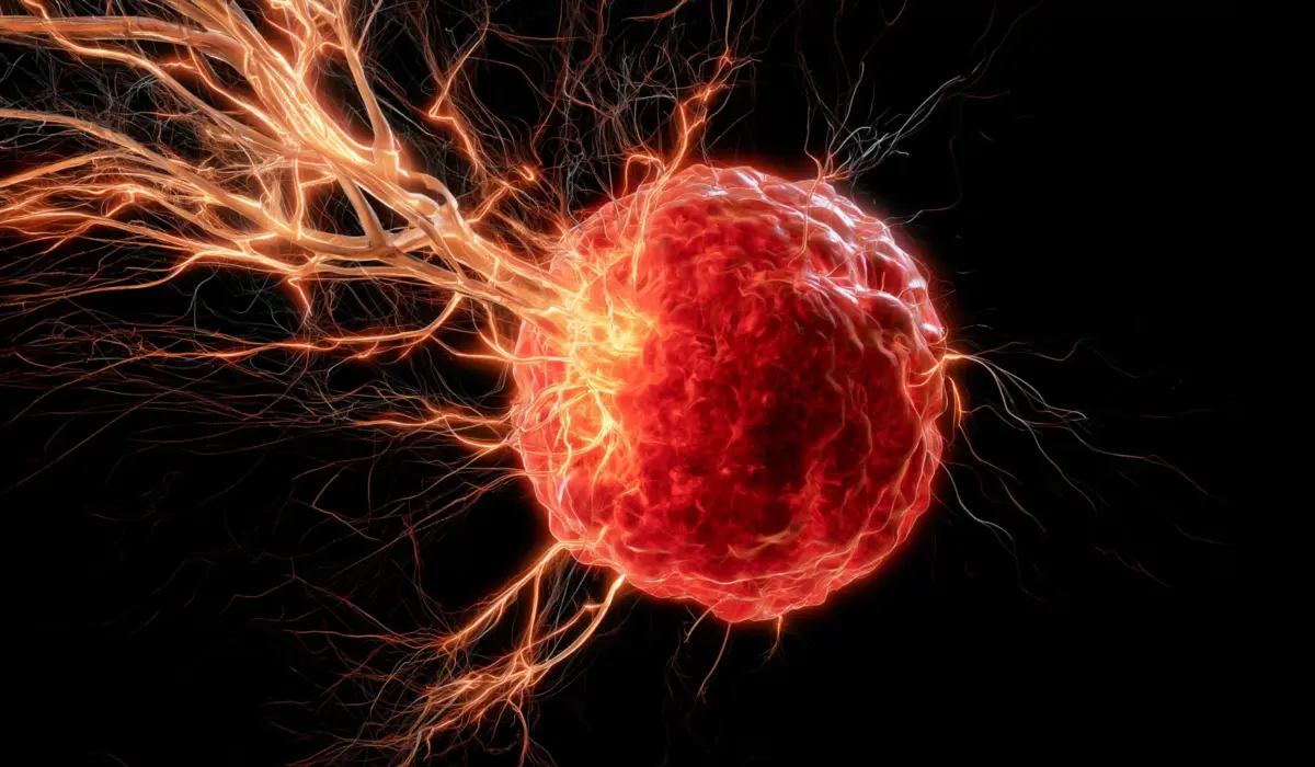
In the relentless battle against oncology’s most formidable adversaries, researchers have long grappled with an insidious biological paradox: the very systems designed to protect the human body can, under the malignant influence of cancer, be subverted into agents of destruction. A groundbreaking study published in the journal Cell Death & Differentiation has laid bare one of the most sophisticated subversions yet discovered. Led by a team of scientists at the University of Oklahoma (OU), the new research reveals how an exceptionally aggressive form of breast cancer manipulates the immune system to weave intricate nerve networks directly into tumors, establishing a hospitable microenvironment that drives cancer progression and treatment resistance.
The investigation focuses on triple-negative breast cancer (TNBC), a notoriously difficult-to-treat subtype that lacks the three major receptors targeted by standard hormonal and targeted therapies: estrogen receptors, progesterone receptors, and human epidermal growth factor receptor 2 (HER2). Because clinicians have fewer targeted options for TNBC, patient outcomes are frequently poor compared to other breast cancer variants. The OU research team has uncovered a critical missing link in understanding how this malignancy thrives: the mechanism by which solid tumors become heavily innervated.
For years, pathologists have observed that many solid tumors contain dense networks of nerves, but the precise etiology of these neural invasions remained an enigma. How do peripheral nerves cross the physiological boundary to infiltrate a growing mass of cancer cells? The OU study answers this question by demonstrating that TNBC tumors recruit specialized immune cells—macrophages—and reprogram them to secrete a neurotrophic protein. This protein acts as a molecular beacon, coaxing nearby nerves to extend their axons directly into the tumor core.
Crucially, this discovery does more than merely map a novel biological pathway; it unveils an entirely unexploited therapeutic vulnerability. When the researchers intervened in pre-clinical models by blocking this specific signaling axis using an already FDA-approved pharmaceutical agent, nerve infiltration ceased, and tumor growth plummeted. Furthermore, analysis of clinical data from human patients corroborated the pre-clinical findings, revealing that elevated levels of these hijacked immune cells and their associated neurotrophic factors directly correlate with diminished survival rates.
As the medical community looks toward the horizon, these findings herald a paradigm shift in oncology. Rather than focusing exclusively on cytotoxic agents designed to eradicate malignant cells directly, future therapies may intercept the crosstalk between immune cells, nerves, and cancer cells, ultimately restoring the body’s natural anti-tumor immunity.
Detailed Chronology: Unraveling the Neural-Immune Axis in Cancer
The path to this discovery represents a convergence of multiple scientific disciplines, blending immunology, neurobiology, and oncology. To fully appreciate the magnitude of the University of Oklahoma’s findings, it is necessary to examine the chronological progression of how cancer biology has evolved to recognize the intimate relationship between nerves and tumors—a field of study increasingly known as cancer neuroscience.
Decades of Observation: The Enigma of Tumor Innervation
For over a century, observant pathologists noted the occasional presence of nerve fibers within malignant tissues, but these observations were largely treated as histological curiosities or incidental findings. It was not until the late 20th and early 21st centuries that modern cancer biologists began to suspect that nerves were not merely passive bystanders in the tumor microenvironment.
Instead, accumulating evidence suggested that tumors actively recruit nervous system components to facilitate their own survival, utilizing neurotransmitters and neurotrophic factors to stimulate cellular proliferation, evade apoptosis, and orchestrate metastasis. However, a major bottleneck in the field persisted: researchers understood that nerves were present within tumors, but the exact molecular mechanisms driving peripheral nerve axons to migrate into the hostile, oxygen-depleted core of a solid tumor remained poorly understood.
The Breakthrough at the University of Oklahoma
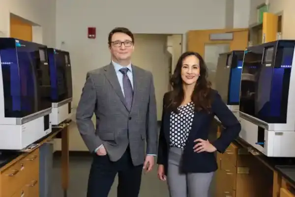
Enter the research team at the University of Oklahoma, spearheaded by Dr. Maureen Cox, an assistant professor in the Department of Microbiology and Immunology at the OU College of Medicine and a research member of the OU Health Stephenson Cancer Center. Focusing their lenses on triple-negative breast cancer, Cox and her colleagues set out to interrogate the cellular mediators responsible for drawing peripheral nerves into the malignant microenvironment.
Through rigorous cellular assays and murine modeling, the team zeroed in on macrophages—a versatile population of white blood cells belonging to the innate immune system. Classically known for their roles in phagocytosis, pathogen clearance, and tissue repair, macrophages are frequently recruited to sites of injury or inflammation. Because a growing tumor is often described as "a wound that never heals," it perpetually emits inflammatory signals that summon macrophages into its fold, where they typically differentiate into tumor-associated macrophages (TAMs).
The OU study revealed a sinister twist in this physiological process. Once ensnared within the triple-negative breast cancer microenvironment, these macrophages are functionally reprogrammed by the tumor. Rather than mounting an anti-tumor immune response or facilitating standard tissue repair, the rogue macrophages begin synthesizing and releasing high levels of brain-derived neurotrophic factor (BDNF).
The Mechanism of Action: BDNF as a Molecular Beacon
Brain-derived neurotrophic factor is a member of the neurotrophin family of growth factors. In a healthy physiological context, BDNF is predominantly synthesized in the central nervous system—particularly in areas like the hippocampus and cortex—where it plays a vital role in supporting the survival, differentiation, and plasticity of neurons.
However, the OU research demonstrated that triple-negative breast cancer exploits this potent neurotrophic signal for pathological ends. The BDNF secreted by the hijacked tumor-associated macrophages diffuses into the surrounding stroma, acting as a powerful chemoattractant and growth cue for nearby peripheral nerve fibers. Sensing the high concentration of BDNF, these nerve axons sprout and extend toward the tumor mass, ultimately penetrating the capsule and establishing a dense intratumoral neural network.
Once embedded within the cancer, these nerves do not sit idle. Emerging hypotheses and correlative data suggest that the newly introduced neural network provides a dual advantage to the malignancy: it may stimulate the angiogenesis required to feed the rapidly dividing cancer cells, and it may actively suppress local immune responses, shielding the tumor from immune-mediated destruction.
Supporting Context & Metrics: The Human Cost and Translational Potential
To contextualize the importance of the OU discovery, it is essential to examine the epidemiological landscape of triple-negative breast cancer, alongside the quantitative metrics that underscore the clinical relevance of the research.
Triple-Negative Breast Cancer: A High-Stakes Clinical Challenge
Breast cancer is not a monolithic disease; it is categorized into distinct molecular subtypes defined by the presence or absence of specific hormonal and growth factor receptors. While hormone receptor-positive and HER2-positive breast cancers have seen dramatic improvements in survival rates over the past two decades due to targeted therapies (such as tamoxifen, aromatase inhibitors, and trastuzumab), triple-negative breast cancer remains stubbornly refractory to these interventions.
- Prevalence: TNBC accounts for approximately 10% to 15% of all breast cancer diagnoses globally.
- Demographic Vulnerability: TNBC disproportionately affects younger women, premenopausal women, and women of African descent, compounding health disparities within oncology.
- Aggressiveness: TNBC is characterized by high histological grade, rapid cellular proliferation, and a heightened propensity for visceral metastasis (particularly to the brain, lungs, and liver).
- Recurrence Rates: Patients with TNBC experience a significantly higher rate of distant recurrence within the first three to five years post-diagnosis compared to other breast cancer subtypes, leaving a narrow window for curative intervention.
Translating Murine Models to Human Pathophysiology
A recurring challenge in oncological research is the translation of discoveries made in animal models to human clinical realities. To bridge this gap, the OU research team did not rely solely on murine experiments; they rigorously interrogated human clinical datasets associated with triple-negative breast cancer.
By analyzing patient tumor samples, the researchers sought to determine whether the biological triad observed in mice—macrophages, BDNF expression, and neural density—mirrored human disease patterns. The correlative data were striking:
- Elevated Biomarkers: Human TNBC tumors characterized by high infiltration of macrophages and elevated expression levels of BDNF exhibited significantly denser nerve networks.
- Prognostic Impact: Patients whose tumors displayed this high-macrophage, high-BDNF, and heavily innervated signature experienced markedly poorer overall survival and progression-free survival rates.
These human metrics validate the pre-clinical hypothesis, confirming that the macrophage-BDNF-nerve axis is not an experimental artifact of murine models, but a genuine driver of human cancer pathophysiology.
Official Statements and Expert Insights
The implications of this study extend far beyond the academic confines of the University of Oklahoma, eliciting commentary from key investigators and setting the stage for future clinical trials.
Dr. Maureen Cox, lead investigator and assistant professor in the Department of Microbiology and Immunology at the OU College of Medicine, as well as a research member of the OU Health Stephenson Cancer Center, articulated the paradoxical nature of the findings during media briefings:
"Macrophages are the critical source for drawing nerves into the tumor. Although macrophages typically play a positive role in the body, they are facilitating a negative function in this scenario of breast cancer."
This quote encapsulates the central tragedy of tumor immunology: physiological mechanisms evolved over millions of years to protect and repair the host are systematically hijacked by malignant clones to construct their own micro-environmental infrastructure.
Addressing the therapeutic potential of intercepting this pathway, Dr. Cox highlighted a particularly exciting avenue for rapid clinical translation:
"It looks really promising that we can use this drug, which is already on the market, to target BDNF. We believe that the nerves are immunosuppressive, so if we can stop the nerves from growing in the first place, maybe we can boost the immune response to help fight the cancer."
The prospect of utilizing an already-marketed pharmaceutical agent—a drug with an established human safety profile—drastically compresses the timeline typically required to move a discovery from a basic science laboratory to human clinical trials. By bypassing early-phase toxicity evaluations that standard de novo drug development demands, the OU team envisions a streamlined path toward clinical implementation.
Furthermore, leadership at the OU Health Stephenson Cancer Center and institutional bodies have underscored the vital importance of sustained research funding in achieving breakthroughs of this caliber. The project’s financial backing reflects a robust coalition of federal and state support, designed to foster translational research that directly benefits patients within Oklahoma and beyond.
Future Outlook: Reversing the Immunosuppressive Tide
With the foundational mechanics of the macrophage-BDNF-nerve axis mapped out in triple-negative breast cancer, the University of Oklahoma research team is already looking toward the next phase of scientific inquiry and clinical application.
Expanding the Therapeutic Target: Repurposing Approved Drugs
The immediate translational goal is the clinical testing of BDNF-targeting interventions in human patients. Because pharmacological agents capable of blocking BDNF or its high-affinity receptor, Tropomyosin receptor kinase B (TrkB), already exist—having been investigated primarily in neurological and psychiatric contexts—repurposing these medications for oncology represents a low-risk, high-reward strategy.
In their pre-clinical murine trials, administering a drug that blocks BDNF signaling yielded a dual benefit:
- Structural Interception: Nerve fibers were successfully prevented from invading the tumor microenvironment.
- Tumor Suppression: Overall tumor growth was significantly curtailed.
Dr. Cox and her colleagues hypothesize that cutting off the neural supply lines does more than starve the tumor of structural support; it fundamentally alters the local immune landscape. If, as current evidence suggests, the infiltrated nerves exert an immunosuppressive effect—actively dampening the ability of T-cells and natural killer cells to recognize and destroy malignant cells—then preventing nerve growth could lift this molecular brake, transforming "cold" tumors (which evade the immune system) into "hot" tumors (which are actively infiltrated and attacked by immune cells).
Uncovering the Mechanics of Metastasis
Beyond therapeutic intervention, the OU team is actively investigating the precise mechanical contributions of nerves to tumor progression. Two primary hypotheses are currently under investigation:
- Vascular Stimulation: Do the invading nerves secrete or stimulate angiogenic factors (such as VEGF) that encourage the formation of new blood vessels, thereby supplying the rapidly growing tumor with the oxygen and glucose required to sustain high metabolic rates?
- Neural Highways for Metastasis: Do cancer cells utilize the physical architecture of nerve fibers as physical migration tracks—a process known as perineural invasion—to escape the primary tumor site, enter the peripheral nervous system or circulatory pathways, and seed distant metastatic colonies in vital organs?
Broadening the Scope: High-Grade Ovarian Cancer
The implications of this research are not restricted to breast cancer. Encouraged by their findings in TNBC, Dr. Cox’s laboratory is already planning to test the same intervention strategy in high-grade ovarian cancer. Ovarian malignancy shares several key features with triple-negative breast cancer: it is notoriously aggressive, frequently diagnosed at an advanced stage, difficult to treat with standard regimens, and characterized by extensive peritoneal innervation and immune cell infiltration.
If blocking BDNF signaling proves equally efficacious in halting nerve infiltration and tumor growth in ovarian cancer models, the therapeutic framework established by the University of Oklahoma could find broad utility across multiple solid tumor types that rely on neural hijacking.
The Ultimate Goal: Restoring Natural Immunity
Ultimately, the vision guiding the OU research team is deceptively simple in its biological ambition, even if breathtaking in its complexity: to restore the patient’s intrinsic capacity for anti-tumor immunity.
By systematically dismantling the protective infrastructure that cancers construct—whether by blocking the immune cells that recruit nerves, neutralizing neurotrophic factors like BDNF, or severing the communication lines between the nervous and immune systems—modern medicine is stepping closer to a future where the body’s own defense mechanisms can reliably recognize, reject, and ultimately eradicate malignancies.
Research Support and Funding Acknowledgments
The groundbreaking research conducted at the University of Oklahoma was made possible through the generous support of federal and state funding agencies committed to advancing biomedical science and oncology care:
- National Institute of General Medical Sciences (NIGMS) of the National Institutes of Health (NIH): Supported through award numbers P20GM103447 and P20GM103639.
- Oklahoma’s Tobacco Settlement Endowment Trust (TSET): A primary funder of the Stephenson Cancer Center and the TSET Health Promotion Research Center at the University of Oklahoma, providing vital infrastructure and operational support.
- Institutional Development Award (IDeA): Supported through the Oklahoma Shared Clinical and Translational Resources via grant number U54GM104938 from the National Institute of General Medical Sciences, facilitating collaborative translational research across regional and national networks.