







Executive Overview
Aging has long been recognized as the single greatest risk factor for neurodegenerative conditions like Alzheimer’s disease and various forms of dementia, yet the precise biological triggers that transition a healthy brain into a vulnerable one have remained elusive. Now, a landmark study funded by the National Institutes of Health (NIH) has exposed a profound, previously hidden transformation in the human brain’s immune landscape.
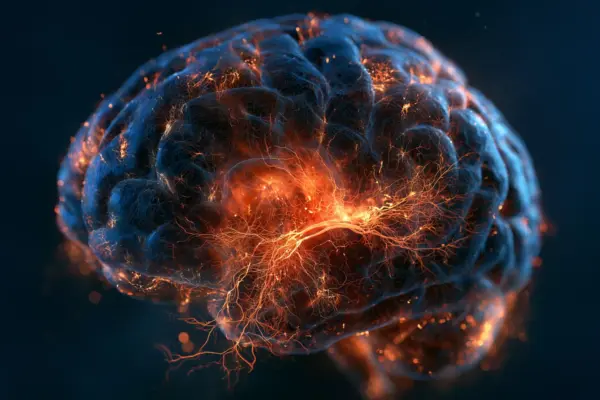
Focusing on the hippocampus—the fundamental clearinghouse for learning, spatial navigation, and memory consolidation—researchers have discovered that a major immune remodeling process quietly begins during midlife.
For decades, neuroscientists operated under the dogma that microglia, the brain’s primary resident immune cells, formed during embryonic development and faithfully maintained themselves in situ throughout an individual’s lifetime through continuous self-renewal. This new research shatters that long-standing assumption. By deploying cutting-edge single-cell multiomic technologies, an interdisciplinary team of scientists from the University of California, San Diego (UCSD), the New York Genome Center, and the University of California, Irvine (UCI), discovered that resident microglia gradually diminish between the ages of 50 and 75.
Crucially, these departing cells are not simply replaced by younger microglia; instead, they are superseded by foreign-acting cells bearing aggressive inflammatory signals and biological signatures strikingly similar to immune cells originating in peripheral blood.
This subtle yet structural immune takeover during middle age may finally decode the long-standing mystery of why chronic, low-grade brain inflammation accompanies biological aging and accelerates cognitive decline. By mapping the epigenetic footprints and three-dimensional nuclear architecture of thousands of individual brain cells, the research team has opened an entirely new window into neuro-immunology.
Published as part of a prestigious compendium of studies in Science and Science Advances backed by the NIH Common Fund’s 4D Nucleome (4DN) program, these findings not only rewrite foundational neuroscience textbooks but also establish a clear molecular target for future therapeutics aimed at preserving cognitive longevity.
Detailed Chronology: Unraveling the Cellular Shift
To comprehend the magnitude of this discovery, it is necessary to trace how the research team systematically dismantled decades of neurological assumptions using unprecedented technological precision.
The Postmortem Cartography of Aging
The investigative journey began with human tissue. Recognizing that animal models often fail to replicate the complex, decades-long trajectory of human brain aging, the researchers turned to postmortem hippocampal tissue samples. Sourced meticulously from 40 neurologically healthy adult donors ranging dramatically in age from 20 to 95 years old, the tissue bank served as a chronological map of the human lifespan.
Using high-resolution single-cell genomic methods, the team peeled back the microscopic layers of the hippocampus. They analyzed not merely a broad cross-section of tissue, but individual cells one by one, cataloging their genetic activity, molecular markers, and structural configurations.
The Midlife Tipping Point (Ages 50–75)
As the researchers mapped the genomic data across the age spectrum, a clear and alarming pattern emerged. In young and early-middle-aged adults, the hippocampal microglial population remained relatively stable, characterized by homeostatic gene expression patterns designed to clean up cellular debris, prune unnecessary synapses, and maintain neural health without triggering damaging inflammation.
However, a dramatic shift materialized precisely within the midlife corridor, spanning roughly from age 50 to age 75. During this developmental window, the population density of resident microglia began a steady, progressive decline.
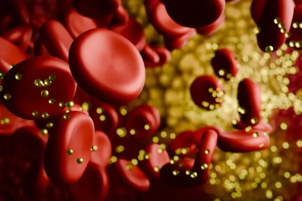
Simultaneously, a second population of cells began to populate the hippocampal landscape. These incoming cells were molecular interlopers: they exhibited heightened inflammatory profiles and shared genetic signatures with peripheral immune cells that normally patrol the body’s circulatory system outside the central nervous system. This revelation suggests that as humans enter their fifties and sixties, the brain’s immune firewall begins to buckle, allowing systemic immune elements to infiltrate and permanently alter the brain’s native microenvironment.
Challenging Biological Dogma
This discovery directly challenges the central tenet of neuro-immunology that has persisted for generations. Previously, it was believed that the blood-brain barrier effectively quarantined the central nervous system, ensuring that resident microglia were self-sustaining entities isolated from the systemic immune system.
The new single-cell data demonstrates that this isolation breaks down far earlier and more comprehensively than previously understood. The midlife transition of the hippocampal immune ecosystem reveals that the brain’s aging process is not a passive fading of faculties, but an active, inflammatory cellular remodeling event.
Supporting Context & Metrics: Advanced Tools and the Blood-Brain Barrier
The success of this study hinged on moving beyond traditional genetic sequencing. Historically, scientists relied heavily on transcriptomics—measuring RNA expression (gene activity)—to understand what a cell was doing. While powerful, RNA expression is transient; it tells researchers what a cell’s current job is, but it often obscures a cell’s lineage and developmental origin.
Decoding the Epigenome and 3D Genome Architecture
To overcome this limitation, the UCSD-led team combined standard RNA measurements with advanced multiomic methods that map the epigenome (chemical modifications to DNA and histone proteins that control gene accessibility) and the three-dimensional structural organization of the genome itself within the nucleus.
- Epigenetic Signatures as Historical Records: While gene expression can fluctuate rapidly based on environmental stress or daily rhythms, epigenetic modifications act as long-term molecular archives. They preserve permanent markers of where a cell originated and how its lineage evolved over decades.
- 3D Nuclear Organization: Supported by the NIH Common Fund’s 4D Nucleome program, the researchers tracked how the physical folding of chromosomes inside the cell nucleus changes over time.
Across virtually all major brain cell types, the aging process was accompanied by widespread, highly coordinated structural disruptions within the genome. These progressive structural breakdowns were directly correlated with shifts in gene regulation and cellular identity. By linking 3D nuclear folding to cell identity, the researchers identified a fundamental, structural feature of human brain aging.
Compromised Defenses: The Blood-Brain Barrier
Compounding the microglial shift, the study also uncovered distinct signs of age-related degradation in the specialized cells responsible for maintaining the blood-brain barrier (BBB). The BBB is a highly selective semipermeable border of endothelial cells, astrocyte end-feet, and pericytes that prevents circulating pathogens, toxins, and peripheral immune cells from indiscriminately entering the sensitive neural tissue of the brain and spinal cord.
The new data reveals that as humans age, the structural integrity of the cells supporting the blood-brain barrier experiences a progressive decline. This structural weakening likely serves as the physical gateway that permits peripheral immune cells to cross into the hippocampus, replacing the dying resident microglia and establishing a permanent, inflammation-prone colony within the brain’s memory center. This convergence of a failing barrier and infiltrating immune cells creates a self-perpetuating loop of chronic neuroinflammation—a hallmark pathology observed in Alzheimer’s disease and other forms of age-related dementia.
Official Statements and Expert Perspectives
The profound implications of these findings have drawn widespread acclaim from leadership across the participating institutions and the National Institutes of Health, underscoring the study’s potential to redirect the future of neurodegenerative research.
Dr. Richard Hodes, director of the National Institute on Aging (NIA) at the NIH, emphasized the critical gap this research fills in our understanding of cognitive decline:
"Aging is the single largest risk factor for dementia, but our understanding of how it drives disease is still incomplete. This previously hidden microglial shift, now uncovered by innovations in technology and thinking, may be an important clue to help us complete the puzzle."
Highlighting the methodological breakthroughs that made this discovery possible, Dr. Nathan Zemke, director of single-cell genomics at the UC San Diego Center for Epigenomics and first author of the study, explained the limitations of older techniques:
"Gene expression tells us what a cell is doing today, but epigenetic signatures preserve information about where a cell came from. By combining these approaches, we uncovered a major shift in the identity and lineage of immune cells in the aging human brain’s immune cells that gene expression data alone would not have revealed."
Dr. Bing Ren, scientific director and CEO of the New York Genome Center, professor of genetics and development at Columbia University, and a corresponding author of the study, pointed to the broader structural implications of the data:
"The progressive structural disruptions were closely linked to shifts in gene regulation and cell identity, potentially revealing a fundamental feature of aging in the human brain."
Looking toward translational applications and therapeutic development, Dr. Xiangmin Xu, professor and director of the Center for Neural Circuit Mapping at UC Irvine and a corresponding author of the study, noted the potential for future medical interventions:
"Understanding these cellular transitions may provide new opportunities to develop interventions that preserve brain function and reduce vulnerability to neurodegenerative disease."
Future Outlook: Implications for Alzheimer’s and Dementia Interventions
As the scientific community digests the weight of these findings, the research roadmap moving forward is exceptionally clear. The identification of midlife immune remodeling in the hippocampus opens up entirely new frontiers for preventative medicine and geriatric neurology.
Immediate Research Objectives
- Causation vs. Correlation: Future studies will investigate the precise molecular mechanisms that trigger the death and clearance of resident microglia between the ages of 50 and 75. Researchers must determine whether microglial loss is an autonomous cellular aging process or a direct consequence of environmental and vascular stressors.
- Mapping Disease Vulnerability: Scientists will actively examine whether the newly identified transition from resident microglia to inflammation-prone peripheral immune cells directly accelerates the onset and pathology of Alzheimer’s disease, tauopathies, and vascular dementias.
- Cross-Tissue Comparisons: Building on the foundational work funded by NIH grants (such as NIA grants R01AG067153 and R01AG082127, alongside the 4D Nucleome program grant 1U01DA052769), researchers will expand single-cell epigenetic mapping to other brain regions beyond the hippocampus—such as the prefrontal cortex and the amygdala—to determine whether immune remodeling occurs uniformly across the entire central nervous system or is uniquely concentrated in memory-processing centers.
The Dawn of Preventive Neuro-Immunology
For decades, pharmacological efforts to treat Alzheimer’s disease have focused largely on clearing amyloid-beta plaques and tau tangles after clinical symptoms of cognitive impairment have already manifested. Unfortunately, many of these late-stage clinical trials have yielded disappointing results, suggesting that intervening after neurodegeneration is well underway may be akin to locking the barn door after the horse has bolted.
The revelation that the brain’s immune system undergoes a radical, inflammatory transformation during midlife—long before clinical dementia presents—suggests a paradigm shift toward preventative intervention. By identifying biomarkers of this midlife microglial shift and blood-brain barrier degradation, clinicians of the future may be able to detect the early cracks in the brain’s immune firewall decades before memory loss begins.
Ultimately, by targeting the epigenetic drivers of structural genome decay and developing pharmacological agents that prevent the infiltration of inflammatory peripheral immune cells into the hippocampus, medical science may soon possess the tools required to interrupt the aging process itself—preserving cognitive vitality and shielding aging populations from the devastating toll of neurodegenerative disease.