







Executive Overview
The diagnostic and therapeutic landscape of bladder cancer is undergoing a profound paradigm shift. Pathologists are no longer tasked simply with identifying the presence of urothelial carcinoma; instead, they must act as specialized molecular and morphologic detectives. Recognizing, classifying, and quantifying histologic subtypes and divergent differentiation has transitioned from an academic exercise to a critical clinical necessity. Accurate subtyping is now recognized as a primary determinant of patient risk stratification, directly influencing the choice between conservative bladder-sparing therapies and radical, life-altering surgical interventions.
According to insights shared by Francesca Khani, MD, associate professor of pathology and urology at Weill Cornell Medicine, during a College of American Pathologists (CAP) session, the morphological diversity of urothelial carcinoma presents a formidable diagnostic challenge. These tumors frequently mimic either benign, inflammatory processes or metastatic malignancies from other primary organs. With the World Health Organization (WHO) updating its classification standards to replace the term "variant" with "subtype," pathologists must employ sophisticated immunohistochemical (IHC) panels and maintain a high index of clinical suspicion to avoid catastrophic misdiagnoses.
This investigative report explores the clinical necessity of subtyping, details three illustrative case studies that highlight common diagnostic pitfalls, dissects the updated WHO taxonomic framework, and outlines the direct therapeutic and prognostic implications of these critical pathologic distinctions.
Detailed Chronology of Diagnostic Pitfalls: Three Critical Case Studies
To understand the high stakes of subtyping in urological pathology, one must examine the clinical realities of the frozen-section suite and the sign-out bench. Dr. Khani presented three cases that demonstrate how easily aggressive malignancies can masquerade as benign conditions or secondary cancers—and vice versa.
Case 1: The Plasmacytoid Masquerader
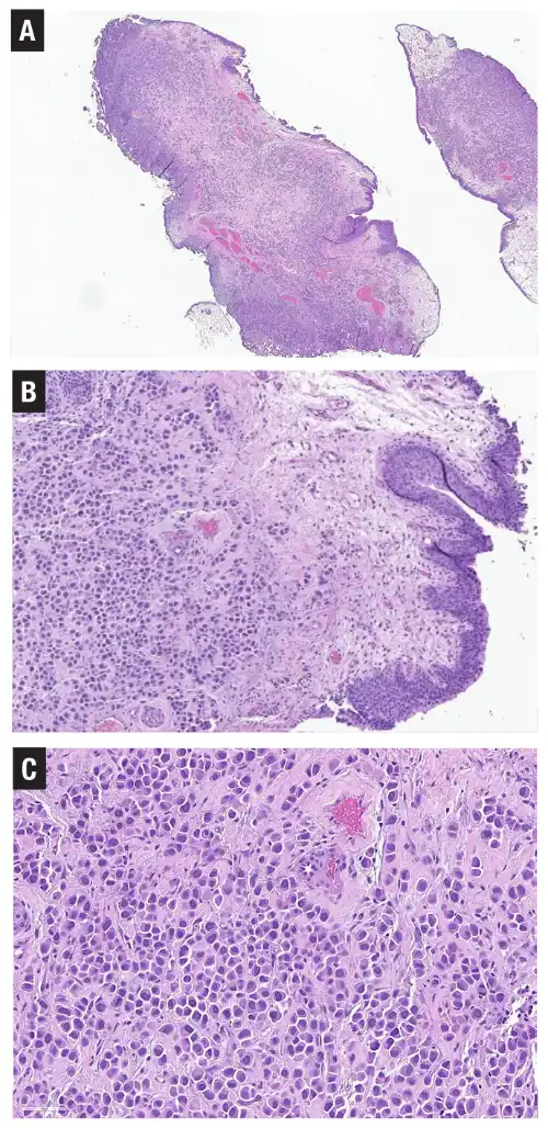
A 73-year-old woman presented to her urologist with persistent lower urinary tract symptoms. A cystoscopic evaluation revealed a diffuse, cobblestone appearance of the entire bladder mucosa, but no discrete, exophytic masses were identified. Operating on the clinical assumption that this represented cystitis cystica et glandularis, the clinical team initially opted not to perform a biopsy, treating the patient instead with multiple courses of antibiotics for a presumed urinary tract infection.
When her symptoms failed to resolve, a repeat cystoscopy and subsequent transurethral resection of bladder tumor (TURBT) were performed. At low magnification, the biopsy specimen revealed a dense, diffuse infiltrate confined to the lamina propria, closely resembling a chronic, non-specific inflammatory process.
[Low Power: Chronic Inflammatory Mimic]
       │
       ├─► [High Power: Discohesive, Single Cells & Linear Files]
       │
       └─► [Highest Power: Eccentric Nuclei (Plasmacytoid Morphology)]
                 │
                 └─► Differential Diagnostic Workup:
                           ├─ Plasmacytoid Urothelial Carcinoma (PUC)
                           ├─ Metastatic Lobular Breast Carcinoma
                           ├─ Signet-Ring / Diffuse GI Primary
                           ├─ Lymphoma / Plasmacytoma
                           └─ Melanoma / Sarcoma
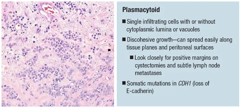
Under higher magnification, however, the cells within the lamina propria demonstrated a highly infiltrative, discohesive growth pattern. The cells grew as single, dispersed units and short, linear cords beneath a completely benign, intact overlying urothelium. At the highest magnification, the neoplastic cells exhibited a classic plasmacytoid appearance: abundant, eosinophilic cytoplasm with eccentrically placed, hyperchromatic nuclei, mimicking benign plasma cells.
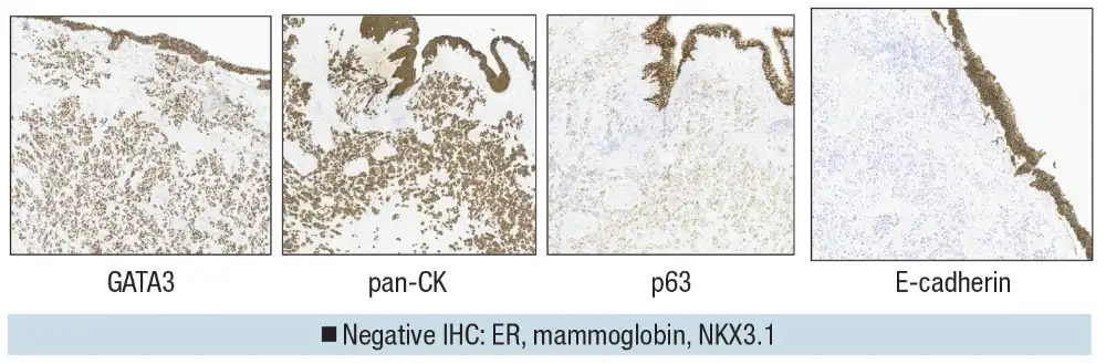
To establish a definitive diagnosis of plasmacytoid urothelial carcinoma (PUC) and rule out key differentials—such as metastatic lobular breast carcinoma, diffuse-type gastric cancer, melanoma, and lymphoma—Dr. Khani’s team deployed a targeted immunohistochemical panel:
- GATA3 and Pan-Cytokeratin (pan-CK): Diffusely positive, confirming an epithelial malignancy and ruling out lymphoma, plasmacytoma, and melanoma.
- p63: Weakly but diffusely positive, pointing toward a urothelial origin and effectively ruling out metastatic lobular breast cancer, which is typically p63-negative.
- E-cadherin: Completely negative, showing a total loss of membranous staining. While E-cadherin loss is the hallmark of lobular breast carcinoma, it is also a well-documented molecular feature of PUC due to somatic mutations in the CDH1 gene.
- NKX3.1: Negative. Though traditionally a prostate-specific marker, NKX3.1 is expressed in a subset of pleomorphic lobular breast carcinomas; its negativity helped further exclude a breast primary.
Through this meticulous IHC profiling, the diagnosis of pure plasmacytoid urothelial carcinoma was confirmed, sparing the patient further ineffective antibiotic treatments and initiating an aggressive oncological intervention.
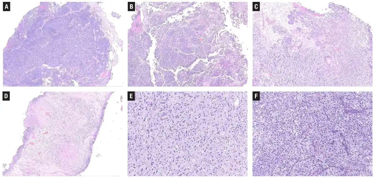
Case 2: The Heterogeneous Prostatic Intruder
A 76-year-old man presented with a bladder mass following a urine cytology evaluation that was read as positive for high-grade urothelial carcinoma. The subsequent TURBT specimen revealed a highly complex, heterogeneous tumor.
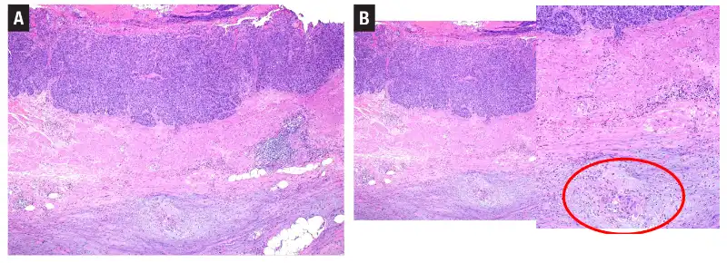
[TURBT Specimen: Complex Heterogeneity]
       ├─► Area A: Nested, inverted growth (mimicking nested UC)
       ├─► Area B: Discohesive, single-cell infiltration of muscularis propria
       ├─► Area C: Cytoplasmic clearing (mimicking clear cell UC)
       └─► Area D: Surface nests (benign von Brunn nests)
The tumor demonstrated several distinct morphological patterns:
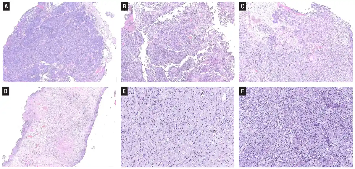
- Solid sheets of cells with nested and inverted growth patterns.
- Focal areas with a hint of papillary architecture supported by delicate fibrovascular cores.
- Regions of highly discohesive, single-cell infiltration deep within the muscularis propria.
- Pockets of marked cytoplasmic clearing, raising suspicion for a clear cell (glycogen-rich) subtype.
While the cytological atypia and deep muscle invasion initially strongly suggested a high-grade, multi-subtype urothelial carcinoma, Dr. Khani noted a critical diagnostic clue: there was no identifiable high-grade urothelial carcinoma in situ (CIS) in the adjacent mucosa, and some paucicellular areas featured remarkably bland nuclei despite the invasive pattern.
Immunohistochemical Resolution:
       ├─► Tumor Cells: NKX3.1 [Strong/Diffuse Positive] ──► Prostatic Adenocarcinoma
       └─► Surface Nests: GATA3 [Positive] / NKX3.1 [Negative] ──► Benign von Brunn Nests
An IHC workup revealed that the entire invasive tumor was strongly and diffusely positive for NKX3.1, while GATA3 was entirely negative within the invasive component. Conversely, the well-circumscribed nested structures near the surface were GATA3-positive and NKX3.1-negative, identifying them as benign, reactive von Brunn nests caught in the tumor’s periphery.
The final diagnosis was metastatic prostatic adenocarcinoma showing treatment-related artifacts. The patient had undergone prior, undisclosed androgen deprivation therapy (hormone therapy), which had altered the tumor’s morphology to mimic the discohesive and clear-cell patterns of aggressive urothelial subtypes. This case highlighted the danger of relying solely on cytological clearance and the critical importance of obtaining a complete clinical history.
Case 3: The Paraganglioma Trap
A 77-year-old woman was found to have an incidental, 1.0-cm enhancing mass located in the dome of her bladder during routine abdominal imaging. A transurethral resection was performed to excise the lesion.
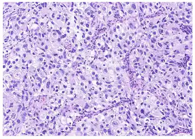
Histological examination of the resection specimen revealed a highly infiltrative, nested architectural pattern that appeared to invade deep into the detrusor muscle. At higher magnification, the tumor cells grew in organoid, sheet-like arrangements. The individual cells displayed cytologically bland, round nuclei with classic "salt-and-pepper" chromatin and abundant, amphophilic-to-eosinophilic cytoplasm, closely mimicking the nested subtype of urothelial carcinoma or a neuroendocrine-differentiated variant.
[Infiltrative Dome Mass] ──► GATA3 (+) & Synaptophysin (+)
                                    │
                         [Diagnostic Fork]
                                    │
         ┌──────────────────────────┴──────────────────────────┐
         ▼                                                     ▼
   Pan-CK (+) / p63 (+)                                  Pan-CK (-) / p63 (-)
[Urothelial Carcinoma]                                    [Paraganglioma]
                                                               (Confirmed)
Initial IHC staining yielded highly confusing, discordant results: the tumor cells were strongly positive for both GATA3 (a primary marker for urothelial differentiation) and synaptophysin (a neuroendocrine marker). GATA3 expression is a well-known diagnostic pitfall in non-urothelial tissues, as it is also consistently expressed in paragangliomas.
To resolve this diagnostic dilemma, Dr. Khani’s team stained the tissue for cytokeratins and p63:
- Pan-cytokeratin (pan-CK): Completely negative.
- p63: Completely negative.
Because a true urothelial carcinoma—even one with extensive neuroendocrine differentiation—retains cytokeratin expression, the complete absence of pan-CK and p63, coupled with GATA3 and synaptophysin positivity, confirmed a diagnosis of benign bladder paraganglioma. Recognizing this mimic prevented the patient from undergoing an unnecessary, highly radical cystectomy.
Supporting Context, Taxonomy, and Diagnostic Metrics
To standardize the reporting of these complex tumors, the World Health Organization (WHO) updated its classification of urinary tract tumors in 2022. A key change was the transition from the term "variant histology" to "histologic subtype" for invasive urothelial carcinomas. This change was designed to avoid clinical confusion with molecular "variants" (i.e., genomic alterations) identified through next-generation sequencing.
Divergent Differentiation vs. Histologic Subtype
As detailed by pathologists Sarah E. Wobker, MD, and colleagues, the distinction between these two terms is clinically important, though they frequently overlap within the same tumor:
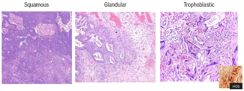
- Divergent Differentiation: Refers to tumor cells that have transdifferentiated to show distinct morphological features of a completely non-urothelial lineage, such as squamous, glandular, or trophoblastic differentiation.
- Histologic Subtype: Represents urothelial cells that have adopted a non-conventional, highly distinct architectural growth pattern (e.g., nested, micropapillary, or tubular) while typically retaining their underlying urothelial cytologic and immunophenotypic profiles.
| Category | Specific Subtypes / Lineages | Morphological & Clinical Mimics | Primary IHC Markers for Diagnosis |
|---|---|---|---|
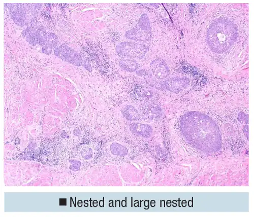
| Mimics of Benign Entities | Nested / Large Nested | von Brunn nests, nephrogenic adenoma | GATA3 (+), p63 (+), Ki-67 (elevated in deep nests) |
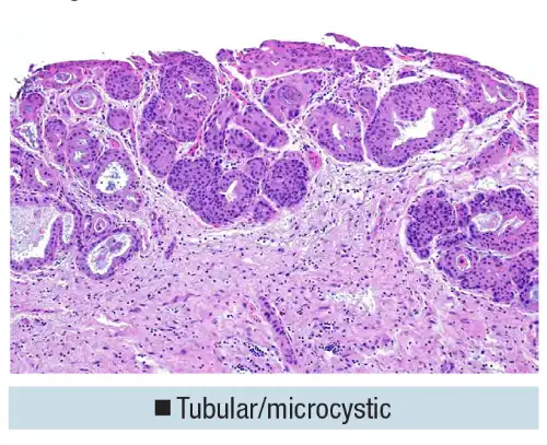
| Tubular & Microcystic | Cystitis cystica et glandularis, nephrogenic adenoma | GATA3 (+), Pax-8 (-) | |
| Mimics of Malignancy | Plasmacytoid | Lobular breast cancer, diffuse gastric cancer, myeloma | GATA3 (+), p63 (+/weak), E-cadherin (lost in 74%) |
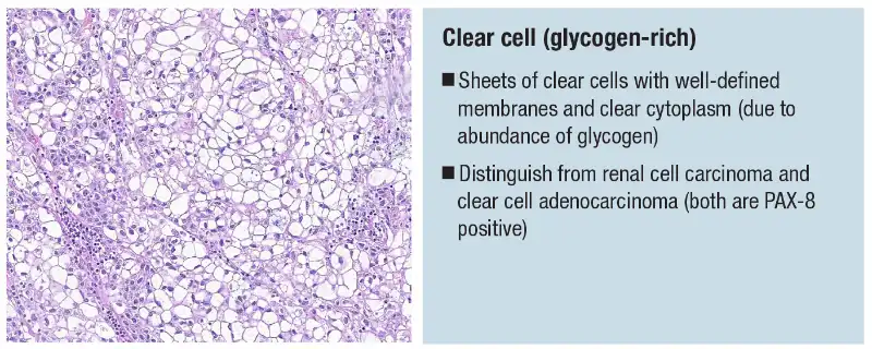
| Clear Cell (Glycogen-Rich) | Renal cell carcinoma (RCC), clear cell adenocarcinoma | GATA3 (+), Pax-8 (-) | |
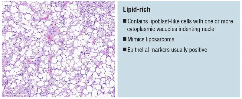
| Lipid-Rich | Liposarcoma, metastatic liposarcoma | GATA3 (+), Pan-CK (+) | |
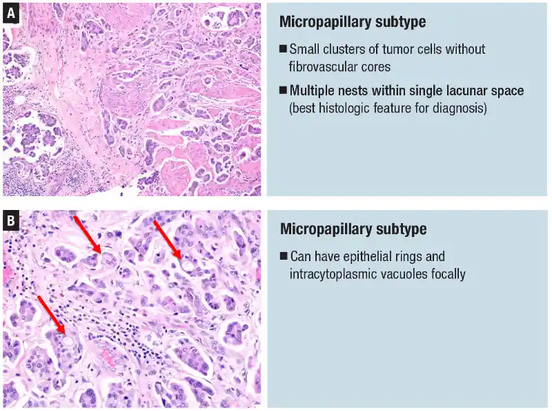
| Aggressive Subtypes | Micropapillary | Retraction artifact, serous ovarian carcinoma | GATA3 (+), HER2 (often overexpressed), MUC1 (inside-out) |
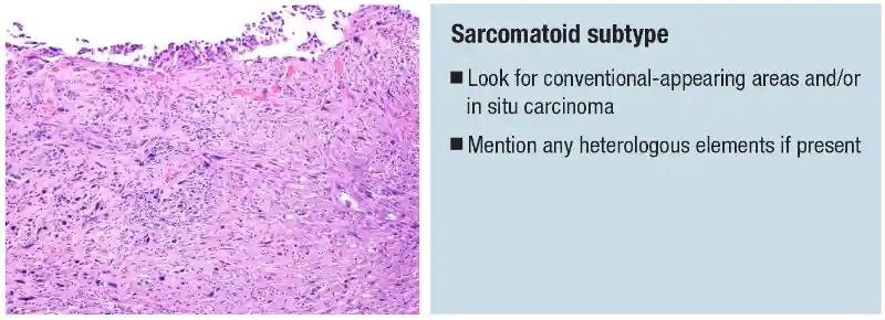
| Sarcomatoid | Primary bladder sarcoma, carcinosarcoma | Pan-CK (+/focal), GATA3 (+/focal), Epithelial membrane antigen (EMA) | |
| Giant Cell / Poorly Diff. | Pleomorphic undifferentiated sarcomas | Pan-CK (+/focal), GATA3 (+/focal) | |
| Favorable Subtype | Lymphoepithelioma-like | Chronic cystitis, lymphoma | Pan-CK (+ in syncytial sheets), PD-L1 (highly enriched) |
Official Statements and Clinical Insights
The clinical importance of identifying these subtypes lies in their direct impact on patient management. In her address at the CAP session, Dr. Khani emphasized that pathologists must not only identify these subtypes but also attempt to quantify their presence within the pathology report.
"Ideally, you want to mention the subtype histologies in pathology reports and quantify the amount," Dr. Khani stated. "If a tumor spreads later on, identifying these subtypes in the primary specimen helps a pathologist in the future recognize it as urothelial when it doesn’t immediately look like it."
This quantification is particularly critical in the setting of non-muscle-invasive bladder cancer (NMIBC), specifically stage pT1 disease. Under the National Comprehensive Cancer Network (NCCN) Guidelines (Version 1.2025) and the American Urological Association (AUA) risk-stratification models, the presence of any aggressive histologic subtype—such as micropapillary, plasmacytoid, or sarcomatoid—automatically places the patient into the "very high-risk" category.
[pT1 Bladder Cancer Diagnosis]
       │
       ├─► Conventional Urothelial Carcinoma ──► Intravesical BCG Therapy (Conservative)
       │
       └─► Aggressive Subtype Present (e.g., Micropapillary, PUC)
                 │
                 └─► [Very High-Risk Stratification] ──► Early Radical Cystectomy (Preferred)
For a standard pT1 conventional urothelial carcinoma, conservative management with transurethral resection followed by intravesical Bacillus Calmette-Guérin (BCG) therapy is often the standard of care. However, if an aggressive subtype is identified, the preferred primary treatment shifts toward early radical cystectomy. Calling out an aggressive subtype can therefore directly trigger a major, life-altering surgical intervention.
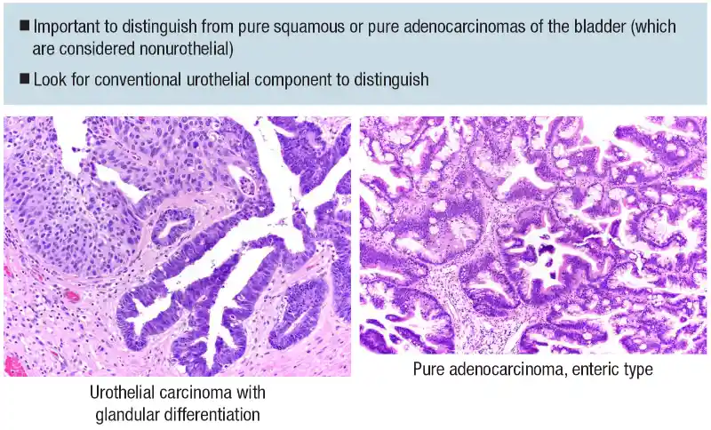
Conversely, distinguishing divergent differentiation from pure non-urothelial malignancies of the bladder is equally critical. For example, a urothelial carcinoma with divergent squamous or glandular differentiation is treated with standard cisplatin-based neoadjuvant chemotherapy (NAC) regimens. However, pure squamous cell carcinoma or pure adenocarcinoma of the bladder shows little to no response to standard NAC, making upfront radical surgery the preferred clinical pathway.
Future Outlook: Precision Medicine and Digital Pathology
As oncology moves further into the era of precision medicine, the integration of morphologic subtyping with molecular profiling is becoming the new standard of care. Future diagnostic workflows are expected to rely heavily on identifying specific therapeutic targets associated with distinct histologic subtypes.
A prime example of this is the micropapillary subtype, which is frequently associated with the amplification and overexpression of the HER2 (ERBB2) oncogene. To address this, a CAP biomarker project team—representing the Cancer, Pathology Electronic Reporting, Immunohistochemistry, Center Guideline, and Molecular Oncology committees—published a comprehensive clinical guidebook in the Archives of Pathology & Laboratory Medicine. This guide establishes standardized protocols for HER2 testing in solid tumors, including urothelial carcinomas, paving the way for targeted therapies like antibody-drug conjugates (ADCs) in patients with aggressive, micropapillary disease.
Similarly, the lymphoepithelioma-like subtype, which is characterized by dense immune cell infiltrates, is highly enriched for PD-L1 expression. This molecular profile makes these tumors exceptionally responsive to immune checkpoint inhibitors, offering a highly effective, bladder-sparing alternative to radical surgery.
Finally, the integration of artificial intelligence (AI) and digital pathology is poised to revolutionize the detection of rare, highly infiltrative subtypes. Machine-learning algorithms are currently being trained to identify the subtle, single-cell discohesive growth patterns of plasmacytoid urothelial carcinoma, which are easily overlooked by the human eye on routine hematoxylin and eosin (H&E) slides. By combining computational precision with the clinical expertise of pathologists, the medical community is moving closer to a future where no aggressive subtype goes unrecognized, ensuring that patients receive the most accurate diagnosis and highly personalized treatment strategies.