




Executive Overview
In a milestone development for computational pathology and cancer diagnostics, an international research team has introduced an advanced deep-learning framework capable of triaging massive three-dimensional (3D) pathology datasets. Published in Nature Biomedical Engineering (2026), the study—led by corresponding author Jonathan T. C. Liu of the University of Washington and Stanford University alongside a consortium of prominent investigators—addresses a critical bottleneck in modern medicine: the inefficiency and potential blind spots of conventional two-dimensional (2D) histopathology.
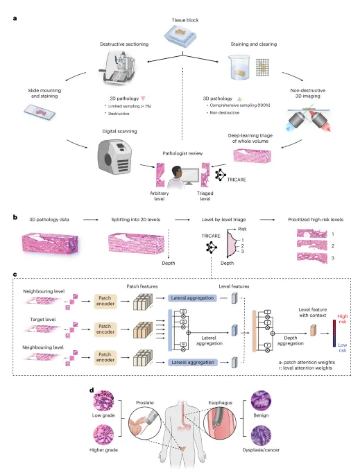
Traditional pathology relies on analyzing thin, isolated 2D tissue slices, a method that leaves up to 99% of a clinical specimen unexamined. This sparse sampling creates vulnerability to diagnostic errors, including the misclassification or undergrading of complex cancers like prostate adenocarcinoma and esophageal dysplasia. The newly developed platform leverages open-top light-sheet (OTLS) microscopy datasets combined with a novel hierarchical multi-instance learning (MIL) architecture known as TRICARE (Transformer-based Range-Integrated Correlated Attention and REpresentation).
By integrating rich structural information across the third dimension, the AI-driven triage system optimizes pathologist workflows. It highlights critical regions of interest across multiple depth levels, ensuring that clinicians can perform comprehensive and efficient evaluations without being overwhelmed by terabytes of volumetric image data. Clinical validations on extensive prostate and esophagus cohorts demonstrated that this technology significantly improves diagnostic accuracy, reduces inter-observer variability, and uncovers high-risk disease features previously missed by conventional 2D histology.
Detailed Chronology & Technological Evolution
The journey toward 3D computational pathology has evolved through distinct technological leaps, culminating in the framework reported in the 2026 Nature Biomedical Engineering study (Gao, G., Yan, R., Song, A.H. et al.).
1. The Limitations of 2D Histopathology
For over a century, diagnostic pathology has depended on slicing tissue blocks into paper-thin sections, mounting them on glass slides, staining them with hematoxylin and eosin (H&E), and examining them under a light microscope. While foundational, this paradigm is fundamentally limited. A standard biopsy core or surgical resection margin consists of a volumetric block of tissue. Reviewing only a few random cross-sections means that focal, high-grade cancer components can easily slip between the sections, resulting in diagnostic discrepancies, patient anxiety, and delayed interventions.
2. Emergence of Optical Sectioning and OTLS Microscopy
To overcome the limitations of physical sectioning, advanced optical imaging modalities—most notably open-top light-sheet (OTLS) microscopy—were developed. OTLS enables rapid, non-destructive, high-resolution imaging of entire cleared tissue blocks, capturing true 3D pathology datasets. However, these volumetric datasets present a paradox: while they eliminate sampling error by capturing the entire specimen, they generate massive amounts of high-resolution image data. Reviewing thousands of optical slices per patient is entirely unfeasible for human pathologists working within standard clinical time constraints.
3. Integrating Deep Learning: The Conception of TRICARE
Recognizing that manual inspection of 3D pathology is impractical, the research collective—featuring contributions from investigators across Stanford University, the University of Washington, the Fred Hutchinson Cancer Research Center, and the University of Pennsylvania—conceived an automated triage solution.
Instead of treating 3D volumes as independent 2D images or forcing raw 3D convolutions that require excessive computational power, the team designed TRICARE. This specialized deep-learning model uses multi-instance learning (MIL) principles to bridge the gap between microscopic patch-level features and macroscopic volumetric risk assessment.
- Patch Encoding & Feature Extraction: The pipeline utilizes state-of-the-art vision foundation models—such as CONCH, UNI, and CTransPath—pretrained on massive collections of whole-slide images (WSIs). These encoders extract rich morphological embeddings from individual tissue patches.
- Range-Integrated Correlated Attention: TRICARE aggregates features not just laterally within a single 2D plane, but also vertically across defined depth ranges ($TRICAREL->D$ or $TRICARED->L$). This allows the network to model spatial-depth correlations, identifying continuous glandular structures, ductal architectures, and epithelial changes across the third dimension.
4. Rigorous Validation and Open Science
The research team rigorously trained and validated the computational models using extensive development cohorts comprising 121 prostate tissue datasets and 334 esophageal tissue datasets. To foster reproducibility and advance the broader computational biology community, the team publicly released all necessary images, annotations, and computational models via Zenodo (DOI: 10.5281/zenodo.20052262), alongside the full 3D pathology datasets for the prostate cohort hosted on The Cancer Imaging Archive (TCIA).
Supporting Context & Quantitative Metrics
The efficacy of the TRICARE framework was evaluated using rigorous cross-validation and comparative ablation analyses against standard 2D aggregation methods. The quantitative findings underscore the superior stability, sensitivity, and clinical utility of 3D-aware AI triage.
Cohort-Level Performance and Aggregation Strategies
When comparing different feature aggregation strategies using CTransPath patch features on the prostate and esophagus development cohorts (evaluated via leave-one-out cross-validation), the 3D-integrated TRICARE approaches consistently outperformed traditional 2D multi-instance learning models.
- Area Under the Curve (AUC): TRICARE configurations achieved superior AUC scores, with tighter 95% confidence intervals calculated via DeLong’s method, indicating robust classification performance across heterogeneous tissue samples.
- Balanced Accuracy and $F_2$ Scores: The framework demonstrated high sensitivity ($F_2$ score optimization), which is vital in oncology to minimize false negatives and ensure that aggressive malignant features are never overlooked.
Ablation Studies and Optimization
Extensive ablation studies were performed to optimize key design parameters:
- Input Formats: The researchers evaluated false-colored (H&E-like) inputs against dual-channel inputs (RGB-format with nuclei in the red channel, eosin in green, and a blank blue channel). Results demonstrated that self-supervised vision models pretrained on H&E whole-slide images performed optimally with H&E-false-colored inputs, with the CONCH foundation model yielding the highest overall performance.
- Lateral Aggregation Architectures: Among tested MIL aggregators (including Attention-Based Multiple Instance Learning [ABMIL], TransMIL, and Double-Tier Feature Distillation MIL [DTFD-MIL]), ABMIL showed superior stability and accuracy.
- Depth Range Tuning: By systematically sweeping the maximum depth range and the number of aggregated levels, the team identified the optimal spatial window. This ensures that the model captures sufficient biological context (such as the continuity of a dysplastic gland) without incorporating irrelevant background noise.
Pathologist Attention Heatmaps and Risk Profiles
Visual inspection of patch attention heatmaps generated by TRICARE revealed remarkable biological alignment:
- Prostate Cohort: High attention scores focused precisely on glandular structures. In higher-grade prostate cancer, the model highlighted crowded and fused glands characteristic of Gleason pattern 4. In low-grade cases, it accurately isolated discrete cancer glands (Gleason pattern 3) and large benign glands.
- Esophagus Cohort: Attention maps concentrated on epithelial surface regions. In dysplastic tissues, the model identified glands characterized by dark, elongated, stratified nuclei with prominent mitoses, whereas mature nondysplastic Barrett’s esophagus surfaces with goblet cells were correctly contextualized.
Furthermore, predicted risk profiles plotted as a function of tissue depth demonstrated that traditional 2D aggregation yields noisy, erratic predictions. In contrast, $TRICARE_L->D$ generated smooth, stable risk curves that aligned closely with ground-truth pathologist evaluations.
Official Statements & Institutional Contributions
The success of this multidisciplinary breakthrough stems from extensive collaboration across leading academic, clinical, and funding institutions.
Principal Investigators and Leadership
- Dr. Jonathan T. C. Liu (University of Washington and Stanford University), the corresponding author and co-inventor of the underlying open-top light-sheet microscopy technologies, emphasized the paradigm-shifting nature of the work: "By transforming how we interrogate volumetric tissue, deep-learning triage bridges the gap between comprehensive 3D imaging and practical clinical review. Pathologists can now navigate complex specimens with unprecedented confidence."
- Dr. Gan Gao, lead computational researcher on the project, noted: "TRICARE successfully addresses the challenge of multi-instance learning across spatial depths. By correlating features vertically and laterally, our model filters out the noise inherent in thick tissue imaging and highlights actionable disease profiles."
Institutional Acknowledgments
The research was made possible through human tissue specimens and clinical samples provided by:
- The Canary Foundation
- The University of Washington
- The Fred Hutchinson Cancer Research Center
- The University of Pennsylvania
Comprehensive Funding Support
The study received substantial backing from federal agencies, private foundations, and industry partnerships:
- National Institutes of Health (NIH): Supported by the National Institute of Diabetes and Digestive and Kidney Diseases (NIDDK) under grant R01DK138948 (J.T.C.L., F.M., W.M.G.), the National Cancer Institute (NCI) under multiple R01, U01, and U54 grants (including R01CA268207, U01CA152756, R01CA220004, U2CCA271902, U54CA163060, and U01CA182940), and the National Heart, Lung, and Blood Institute (NHLBI) under R01HL15127701A1 and R01HL15807101A1 (A.M.).
- Advanced Research Projects Agency for Health (ARPA-H): Contract numbers D24AC00357 (J.T.C.L.) and D25AC00140 (A.M.).
- Department of Defense (DoD): Prostate Cancer Research Program (PCRP) grants W81XWH-18-10358 (J.T.C.L., L.D.T.), W81XWH-14-2-0183 (L.D.T.), and W81XWH-20-1-0851 (A.M., J.T.C.L.).
- Philanthropic and Institutional Funds: The Prevent Cancer Foundation, Cottrell Family Fund, Evergreen Fund, Listwin Foundation, Canary Foundation, GiCaRes, Pacific Northwest Prostate Cancer SPORE P50CA97186, and NSF Graduate Research Fellowship DGE-1762114.
- Industry Collaborations: Sponsored research agreements and consulting ties with Bristol Myers Squibb, AstraZeneca, Takeda Inc., Freenome, Guardant Health, SEngine, and Alpenglow Biosciences, Inc.
(Note: The funding agencies and institutional sponsors bore no editorial control over the study design, execution, or official conclusions, which remain strictly the responsibility of the authors.)
Future Outlook & Clinical Implications
The introduction of deep-learning triage for 3D pathology datasets marks a crucial turning point in diagnostic medicine. As hospitals and diagnostic laboratories increasingly adopt optical clearing and non-destructive 3D imaging technologies, data volume will continue to surge. Frameworks like TRICARE will no longer be optional research tools; they will become essential operational infrastructure in precision oncology.
Clinical Translation and Workflow Integration
In clinical validation trials, examining a larger number of AI-triaged depth levels (e.g., eight levels versus three) enabled pathologists to identify additional high-risk samples that would otherwise have been missed. Furthermore, AI-triaged 3D pathology frequently enabled the accurate "upgrading" of biopsy cases—revealing fused glands or poorly formed structures indicative of aggressive Gleason pattern 4 prostate cancer or esophageal dysplasia that conventional 2D random sections failed to capture.
Future developments will focus on:
- Real-Time Clinical Trials: Expanding multi-center prospective trials to evaluate the impact of TRICARE-triaged workflows on patient treatment planning and oncological outcomes.
- Multimodal AI Integration: Combining 3D histopathological imaging features with genomic, transcriptomic, and clinical metadata to build holistic prognostic models.
- Software Commercialization: Translating these academic pipelines into FDA-cleared software suites that integrate seamlessly with digital pathology viewers and hospital picture archiving and communication systems (PACS).
By transforming terabytes of complex 3D tissue scans into focused, intuitive, and highly accurate diagnostic roadmaps, deep-learning triage promises to eliminate sampling error, empower pathologists, and ultimately deliver safer, more precise diagnoses for cancer patients worldwide.